 Movie
Movie Controller


+ Open data

- Basic information
| Entry | Database: PDB / ID: 6u66 | ||||||
|---|---|---|---|---|---|---|---|
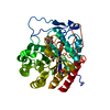
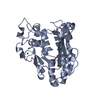
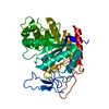
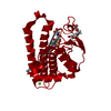
| Title | Structure of the trimeric globular domain of Adiponectin | ||||||
 Components Components | Adiponectin | ||||||
 Keywords Keywords | HORMONE / globular domain | ||||||
| Function / homology |  Function and homology information Function and homology informationnegative regulation of intracellular protein transport / positive regulation of lipid transporter activity / positive regulation of metanephric podocyte development / positive regulation of renal albumin absorption / negative regulation of metanephric mesenchymal cell migration / response to linoleic acid / positive regulation of glycogen (starch) synthase activity / negative regulation of platelet-derived growth factor receptor-alpha signaling pathway / positive regulation of myeloid cell apoptotic process / negative regulation of macrophage differentiation ...negative regulation of intracellular protein transport / positive regulation of lipid transporter activity / positive regulation of metanephric podocyte development / positive regulation of renal albumin absorption / negative regulation of metanephric mesenchymal cell migration / response to linoleic acid / positive regulation of glycogen (starch) synthase activity / negative regulation of platelet-derived growth factor receptor-alpha signaling pathway / positive regulation of myeloid cell apoptotic process / negative regulation of macrophage differentiation / AMPK inhibits chREBP transcriptional activation activity / detection of oxidative stress / positive regulation of signal transduction / negative regulation of granulocyte differentiation / negative regulation of hormone secretion / low-density lipoprotein particle clearance / negative regulation of protein autophosphorylation / sialic acid binding / negative regulation of heterotypic cell-cell adhesion / response to sucrose / negative regulation of vascular associated smooth muscle cell migration / collagen trimer / negative regulation of low-density lipoprotein receptor activity / negative regulation of DNA biosynthetic process / negative regulation of platelet-derived growth factor receptor signaling pathway / positive regulation of monocyte chemotactic protein-1 production / negative regulation of synaptic transmission / positive regulation of cAMP-dependent protein kinase activity / negative regulation of phagocytosis / negative regulation of cold-induced thermogenesis / positive regulation of fatty acid metabolic process / fatty acid beta-oxidation / negative regulation of fat cell differentiation / positive regulation of protein kinase A signaling / fatty acid oxidation / negative regulation of vascular associated smooth muscle cell proliferation / negative regulation of macrophage derived foam cell differentiation / negative regulation of tumor necrosis factor production / regulation of glucose metabolic process / negative regulation of tumor necrosis factor-mediated signaling pathway / positive regulation of cholesterol efflux / response to tumor necrosis factor / negative regulation of gluconeogenesis / response to glucose / brown fat cell differentiation / negative regulation of canonical NF-kappaB signal transduction / cellular response to cAMP / response to glucocorticoid / cellular response to epinephrine stimulus / negative regulation of blood pressure / protein serine/threonine kinase activator activity / negative regulation of cell migration / response to nutrient / response to activity / negative regulation of MAP kinase activity / negative regulation of receptor binding / generation of precursor metabolites and energy / cytokine activity / protein localization to plasma membrane / positive regulation of interleukin-8 production / positive regulation of glucose import / response to bacterium / hormone activity / negative regulation of ERK1 and ERK2 cascade / negative regulation of inflammatory response / Transcriptional regulation of white adipocyte differentiation / circadian rhythm / cellular response to insulin stimulus / glucose metabolic process / positive regulation of peptidyl-tyrosine phosphorylation / cellular response to xenobiotic stimulus / glucose homeostasis / positive regulation of cold-induced thermogenesis / gene expression / response to ethanol / positive regulation of canonical NF-kappaB signal transduction / collagen-containing extracellular matrix / response to hypoxia / positive regulation of protein phosphorylation / signaling receptor binding / negative regulation of DNA-templated transcription / cell surface / endoplasmic reticulum / protein homodimerization activity / extracellular space / extracellular regionSimilarity search - Function | ||||||
| Biological species |  Homo sapiens (human) Homo sapiens (human) | ||||||
| Method | X-RAY DIFFRACTION / SYNCHROTRON / MOLECULAR REPLACEMENT / Resolution: 0.99 Å | ||||||
 Authors Authors | Pascolutti, R. / Kruse, A.C. / Erlandson, S.C. / Burri, D.J. / Zheng, S. | ||||||
| Funding support |  United States, 1items United States, 1items
| ||||||
 Citation Citation | Journal: J.Biol.Chem. / Year: 2020 Title: Mapping and engineering the interaction between adiponectin and T-cadherin. Authors: Pascolutti, R. / Erlandson, S.C. / Burri, D.J. / Zheng, S. / Kruse, A.C. | ||||||
| History |
|
- Structure visualization
| Structure viewer | Molecule: MolmilJmol/JSmol |
|---|
- Downloads & links
-Download
| PDBx/mmCIF format | 6u66.cif.gz | 279.5 KB | Display | PDBx/mmCIF format |
|---|---|---|---|---|
| PDB format | pdb6u66.ent.gz | 230.2 KB | Display | PDB format |
| PDBx/mmJSON format | 6u66.json.gz | Tree view | PDBx/mmJSON format | |
| Others |  Other downloads Other downloads |
-Validation report
| Arichive directory | https://data.pdbj.org/pub/pdb/validation_reports/u6/6u66ftp://data.pdbj.org/pub/pdb/validation_reports/u6/6u66 | HTTPS FTP |
|---|
-Related structure data
| Related structure data | 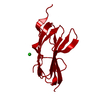 6u6nC 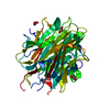 4douS S: Starting model for refinement C: citing same article ( |
|---|---|
| Similar structure data |


-Links
 PDBj
PDBj- Assembly
| Deposited unit | 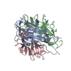
| ||||||||
|---|---|---|---|---|---|---|---|---|---|
| 1 |
| ||||||||
| Unit cell |
|
-Components
| #1: Protein | / 30 kDa adipocyte complement-related protein / Adipocyte complement-related 30 kDa protein / ACRP30 ...30 kDa adipocyte complement-related protein / Adipocyte complement-related 30 kDa protein / ACRP30 / Adipocyte / C1q and collagen domain-containing protein / Adipose most abundant gene transcript 1 protein / apM-1 / Gelatin-binding protein Mass: 16275.028 Da / Num. of mol.: 3 Source method: isolated from a genetically manipulated source Source: (gene. exp.) Homo sapiens (human) / Gene: ADIPOQ, ACDC, ACRP30, APM1, GBP28 / Plasmid: pMAL / Production host:  Escherichia coli BL21 (bacteria) / Strain (production host): BL21 / References: UniProt: Q15848 Escherichia coli BL21 (bacteria) / Strain (production host): BL21 / References: UniProt: Q15848#2: Chemical |   Mass: 40.078 Da / Num. of mol.: 3 / Source method: obtained synthetically / Formula: Ca / Feature type: SUBJECT OF INVESTIGATION Mass: 40.078 Da / Num. of mol.: 3 / Source method: obtained synthetically / Formula: Ca / Feature type: SUBJECT OF INVESTIGATION#3: Chemical | ChemComp-NA / |   Mass: 22.990 Da / Num. of mol.: 1 / Source method: obtained synthetically / Formula: Na / Feature type: SUBJECT OF INVESTIGATION Mass: 22.990 Da / Num. of mol.: 1 / Source method: obtained synthetically / Formula: Na / Feature type: SUBJECT OF INVESTIGATION#4: Water | ChemComp-HOH / | Water Mass: 18.015 Da / Num. of mol.: 537 / Source method: isolated from a natural source / Formula: H2O Mass: 18.015 Da / Num. of mol.: 537 / Source method: isolated from a natural source / Formula: H2OHas ligand of interest | Y | |
|---|
-Experimental details
-Experiment
| Experiment | Method: X-RAY DIFFRACTION / Number of used crystals: 1 |
|---|
- Sample preparation
| Crystal | Density Matthews: 2.01 Å3/Da / Density % sol: 38.67 % |
|---|---|
| Crystal grow | Temperature: 298 K / Method: vapor diffusion, sitting drop / pH: 4.5 Details: 0.2 mM Li2SO4, 0.1 M sodium acetate pH 4.5, 50% PEG 400 PH range: 4-6.5 |
-Data collection
| Diffraction | Mean temperature: 100 K / Serial crystal experiment: N | ||||||||||||||||||||||||||||||||||||||||||||||||||||||||||||||||||||||||||||||||
|---|---|---|---|---|---|---|---|---|---|---|---|---|---|---|---|---|---|---|---|---|---|---|---|---|---|---|---|---|---|---|---|---|---|---|---|---|---|---|---|---|---|---|---|---|---|---|---|---|---|---|---|---|---|---|---|---|---|---|---|---|---|---|---|---|---|---|---|---|---|---|---|---|---|---|---|---|---|---|---|---|---|
| Diffraction source | Source: SYNCHROTRON / Site: APS / Beamline: 23-ID-D / Wavelength: 1.033 Å | ||||||||||||||||||||||||||||||||||||||||||||||||||||||||||||||||||||||||||||||||
| Detector | Type: DECTRIS PILATUS 6M / Detector: PIXEL / Date: Jul 28, 2016 | ||||||||||||||||||||||||||||||||||||||||||||||||||||||||||||||||||||||||||||||||
| Radiation | Monochromator: Double Crystal Si(111) / Protocol: SINGLE WAVELENGTH / Monochromatic (M) / Laue (L): M / Scattering type: x-ray | ||||||||||||||||||||||||||||||||||||||||||||||||||||||||||||||||||||||||||||||||
| Radiation wavelength | Wavelength: 1.033 Å / Relative weight: 1 | ||||||||||||||||||||||||||||||||||||||||||||||||||||||||||||||||||||||||||||||||
| Reflection | Resolution: 0.989→50 Å / Num. obs: 199981 / % possible obs: 95.6 % / Redundancy: 3.95 % / CC1/2: 0.997 / Rmerge(I) obs: 0.134 / Rrim(I) all: 0.155 / Net I/σ(I): 5.01 | ||||||||||||||||||||||||||||||||||||||||||||||||||||||||||||||||||||||||||||||||
| Reflection shell | Diffraction-ID: 1
|
- Processing
| Software |
| ||||||||||||||||||||||||||||||||||||||||||||||||||||||||||||||||||||||||||||||||||||||||||||||||||||||||||||||||||||||||||||||||||||||||||||||||||||||||||||||||||||||||||||||||||||||||||
|---|---|---|---|---|---|---|---|---|---|---|---|---|---|---|---|---|---|---|---|---|---|---|---|---|---|---|---|---|---|---|---|---|---|---|---|---|---|---|---|---|---|---|---|---|---|---|---|---|---|---|---|---|---|---|---|---|---|---|---|---|---|---|---|---|---|---|---|---|---|---|---|---|---|---|---|---|---|---|---|---|---|---|---|---|---|---|---|---|---|---|---|---|---|---|---|---|---|---|---|---|---|---|---|---|---|---|---|---|---|---|---|---|---|---|---|---|---|---|---|---|---|---|---|---|---|---|---|---|---|---|---|---|---|---|---|---|---|---|---|---|---|---|---|---|---|---|---|---|---|---|---|---|---|---|---|---|---|---|---|---|---|---|---|---|---|---|---|---|---|---|---|---|---|---|---|---|---|---|---|---|---|---|---|---|---|---|---|
| Refinement | Method to determine structure: MOLECULAR REPLACEMENT Starting model: 4DOU Resolution: 0.99→40.768 Å / SU ML: 0.16 / Cross valid method: THROUGHOUT / σ(F): 1.96 / Phase error: 24.54
| ||||||||||||||||||||||||||||||||||||||||||||||||||||||||||||||||||||||||||||||||||||||||||||||||||||||||||||||||||||||||||||||||||||||||||||||||||||||||||||||||||||||||||||||||||||||||||
| Solvent computation | Shrinkage radii: 0.9 Å / VDW probe radii: 1.11 Å | ||||||||||||||||||||||||||||||||||||||||||||||||||||||||||||||||||||||||||||||||||||||||||||||||||||||||||||||||||||||||||||||||||||||||||||||||||||||||||||||||||||||||||||||||||||||||||
| Displacement parameters | Biso max: 58.41 Å2 / Biso mean: 14.75 Å2 / Biso min: 5.9 Å2 | ||||||||||||||||||||||||||||||||||||||||||||||||||||||||||||||||||||||||||||||||||||||||||||||||||||||||||||||||||||||||||||||||||||||||||||||||||||||||||||||||||||||||||||||||||||||||||
| Refinement step | Cycle: final / Resolution: 0.99→40.768 Å
| ||||||||||||||||||||||||||||||||||||||||||||||||||||||||||||||||||||||||||||||||||||||||||||||||||||||||||||||||||||||||||||||||||||||||||||||||||||||||||||||||||||||||||||||||||||||||||
| LS refinement shell | Refine-ID: X-RAY DIFFRACTION / Rfactor Rfree error: 0
|
