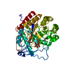
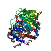
[English] 日本語
Yorodumi
- PDB-6far: Structure of the GH99 endo-alpha-mannanase from Bacteroides xylan... -

+
Open data


ID or keywords:

Loading...

-
Basic information

Entry
Database: PDB / ID: 6far
TitleStructure of the GH99 endo-alpha-mannanase from Bacteroides xylanisolvens in complex with mannose-alpha-1,3-mannoimidazole
ComponentsGlycosyl hydrolase family 71
KeywordsHYDROLASE / complex / GH99
Function / homology
Function and homology information


hydrolase activity, acting on glycosyl bonds / membrane
Similarity search - Function
Glycosyl hydrolase family 99 / Glycosyl hydrolase family 99 / Glycosidases / TIM Barrel / Alpha-Beta Barrel / Alpha Beta
Similarity search - Domain/homology
alpha-D-mannopyranose / Chem-MVL / Glycosyl hydrolase family 71
Similarity search - Component
Biological speciesBacteroides xylanisolvens XB1A (bacteria)
MethodX-RAY DIFFRACTION / SYNCHROTRON / MOLECULAR REPLACEMENT / Resolution: 1.3 Å
AuthorsFernandes, P.Z. / Petricevic, M. / Sobala, L.F. / Davies, G.J. / Williams, S.J.
Funding support United Kingdom, Australia, 3items
OrganizationGrant numberCountry
European Research Council322942 United Kingdom
Australian Research CouncilFT130100103 Australia
Australian Research CouncilDP120101396 Australia
CitationJournal: Chemistry / Year: 2018
Title: Exploration of Strategies for Mechanism-Based Inhibitor Design for Family GH99 endo-alpha-1,2-Mannanases.
Authors: Fernandes, P.Z. / Petricevic, M. / Sobala, L. / Davies, G.J. / Williams, S.J.
History
DepositionDec 16, 2017Deposition site: PDBE / Processing site: PDBE
Revision 1.0Mar 21, 2018Provider: repository / Type: Initial release
Revision 1.1Jun 13, 2018Group: Data collection / Database references / Category: citation
Item: _citation.journal_volume / _citation.page_first ..._citation.journal_volume / _citation.page_first / _citation.page_last / _citation.title
Revision 1.2Jul 29, 2020Group: Data collection / Derived calculations / Structure summary
Category: chem_comp / entity ...chem_comp / entity / pdbx_chem_comp_identifier / pdbx_entity_nonpoly / struct_site / struct_site_gen
Item: _chem_comp.name / _chem_comp.type ..._chem_comp.name / _chem_comp.type / _entity.pdbx_description / _pdbx_entity_nonpoly.name
Description: Carbohydrate remediation / Provider: repository / Type: Remediation
Revision 1.3Feb 3, 2021Group: Data collection / Database references / Structure summary
Category: chem_comp / diffrn_source / pdbx_related_exp_data_set
Item: _chem_comp.pdbx_synonyms / _diffrn_source.pdbx_wavelength_list
Revision 1.4Jan 17, 2024Group: Data collection / Database references / Refinement description
Category: chem_comp_atom / chem_comp_bond ...chem_comp_atom / chem_comp_bond / database_2 / diffrn_radiation_wavelength / pdbx_initial_refinement_model
Item: _database_2.pdbx_DOI / _database_2.pdbx_database_accession / _diffrn_radiation_wavelength.wavelength

-
Structure visualization

Structure viewerMolecule:
MolmilJmol/JSmol

Downloads & links

-
Assembly

Deposited unit
A: Glycosyl hydrolase family 71
hetero molecules


Theoretical massNumber of molelcules
Total (without water)44,3143
Polymers43,9341
Non-polymers3802
Water7,656425
1


  • Idetical with deposited unit
  • defined by author&software
  • Evidence: gel filtration
TypeNameSymmetry operationNumber
identity operation1_555x,y,z1
Buried area310 Å2
ΔGint4 kcal/mol
Surface area13980 Å2
MethodPISA
Unit cell
Length a, b, c (Å)108.619, 108.619, 67.766
Angle α, β, γ (deg.)90.00, 90.00, 90.00
Int Tables number79
Space group name H-MI4
Components on special symmetry positions
IDModelComponents
11A-776-

HOH

-
Components

#1: Protein Glycosyl hydrolase family 71 / Glycosyl hydrolase family 99


Mass: 43933.594 Da / Num. of mol.: 1
Source method: isolated from a genetically manipulated source
Source: (gene. exp.) Bacteroides xylanisolvens XB1A (bacteria)
Gene: BXY_34140 / Plasmid: pET-28a (+) / Production host: Escherichia coli BL21(DE3) (bacteria) / References: UniProt: D6D1V7
#2: Sugar ChemComp-MAN / alpha-D-mannopyranose / alpha-D-mannose / D-mannose / mannose / Mannose


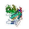
Type: D-saccharide, alpha linking / Mass: 180.156 Da / Num. of mol.: 1
Source method: isolated from a genetically manipulated source
Formula: C6H12O6
IdentifierTypeProgram
DManpaCONDENSED IUPAC CARBOHYDRATE SYMBOLGMML 1.0
a-D-mannopyranoseCOMMON NAMEGMML 1.0
a-D-ManpIUPAC CARBOHYDRATE SYMBOLPDB-CARE 1.0
ManSNFG CARBOHYDRATE SYMBOLGMML 1.0
#3: Chemical ChemComp-MVL / (5R,6R,7S,8R)-5-(HYDROXYMETHYL)-5,6,7,8-TETRAHYDROIMIDAZO[1,2-A]PYRIDINE-6,7,8-TRIOL / Mannoimidazole


Mass: 200.192 Da / Num. of mol.: 1 / Source method: obtained synthetically / Formula: C8H12N2O4 / Feature type: SUBJECT OF INVESTIGATION
#4: Water ChemComp-HOH / water / Water


Mass: 18.015 Da / Num. of mol.: 425 / Source method: isolated from a natural source / Formula: H2O

-
Experimental details

-
Experiment

ExperimentMethod: X-RAY DIFFRACTION / Number of used crystals: 1

-
Sample preparation

CrystalDensity Matthews: 2.27 Å3/Da / Density % sol: 45.93 %
Crystal growTemperature: 292 K / Method: vapor diffusion, hanging drop / pH: 7.4 / Details: 3 M sodium acetate, pH 7.4

-
Data collection

DiffractionMean temperature: 100 K
Diffraction sourceSource: SYNCHROTRON / Site: Diamond / Beamline: I04 / Wavelength: 0.9795 Å
DetectorType: DECTRIS PILATUS 6M / Detector: PIXEL / Date: Jul 5, 2017
RadiationProtocol: SINGLE WAVELENGTH / Monochromatic (M) / Laue (L): M / Scattering type: x-ray
Radiation wavelengthWavelength: 0.9795 Å / Relative weight: 1
ReflectionResolution: 1.3→76.81 Å / Num. obs: 96147 / % possible obs: 99.5 % / Redundancy: 7.5 % / Biso Wilson estimate: 15.83 Å2 / CC1/2: 0.999 / Rmerge(I) obs: 0.054 / Rpim(I) all: 0.02 / Rrim(I) all: 0.057 / Net I/σ(I): 14
Reflection shellResolution: 1.3→1.32 Å / Redundancy: 4.6 % / Rmerge(I) obs: 1.224 / Num. unique obs: 4400 / CC1/2: 0.486 / Rpim(I) all: 0.754 / Rrim(I) all: 1.376 / % possible all: 92.7

-
Processing

Software
NameVersionClassification
REFMAC5.8.0189refinement
DIALS1.6.3-g2108d754-releasedata reduction
Aimless0.5.32data scaling
REFMAC5.8.0158phasing
RefinementMethod to determine structure: MOLECULAR REPLACEMENT
Starting model: 5M17
Resolution: 1.3→76.81 Å / Cor.coef. Fo:Fc: 0.982 / Cor.coef. Fo:Fc free: 0.978 / SU B: 1.758 / SU ML: 0.032 / Cross valid method: THROUGHOUT / ESU R: 0.044 / ESU R Free: 0.043 / Details: HYDROGENS HAVE BEEN ADDED IN THE RIDING POSITIONS
RfactorNum. reflection% reflectionSelection details
Rfree0.16168 4810 5 %RANDOM
Rwork0.13398 ---
obs0.13537 91334 99.43 %-
Solvent computationIon probe radii: 0.8 Å / Shrinkage radii: 0.8 Å / VDW probe radii: 1.2 Å
Displacement parametersBiso mean: 20.958 Å2
Baniso -1Baniso -2Baniso -3
1--0.52 Å20 Å20 Å2
2---0.52 Å20 Å2
3---1.04 Å2
Refinement stepCycle: 1 / Resolution: 1.3→76.81 Å
ProteinNucleic acidLigandSolventTotal
Num. atoms2800 0 25 425 3250
Refine LS restraints
Refine-IDTypeDev idealDev ideal targetNumber
X-RAY DIFFRACTIONr_bond_refined_d0.0110.023327
X-RAY DIFFRACTIONr_bond_other_d0.0020.022950
X-RAY DIFFRACTIONr_angle_refined_deg1.4971.9514618
X-RAY DIFFRACTIONr_angle_other_deg1.00836927
X-RAY DIFFRACTIONr_dihedral_angle_1_deg5.8135453
X-RAY DIFFRACTIONr_dihedral_angle_2_deg36.3223.455165
X-RAY DIFFRACTIONr_dihedral_angle_3_deg13.06115542
X-RAY DIFFRACTIONr_dihedral_angle_4_deg15.1811521
X-RAY DIFFRACTIONr_chiral_restr0.0980.2484
X-RAY DIFFRACTIONr_gen_planes_refined0.0090.0213829
X-RAY DIFFRACTIONr_gen_planes_other0.0020.02758
X-RAY DIFFRACTIONr_nbd_refined
X-RAY DIFFRACTIONr_nbd_other
X-RAY DIFFRACTIONr_nbtor_refined
X-RAY DIFFRACTIONr_nbtor_other
X-RAY DIFFRACTIONr_xyhbond_nbd_refined
X-RAY DIFFRACTIONr_xyhbond_nbd_other
X-RAY DIFFRACTIONr_metal_ion_refined
X-RAY DIFFRACTIONr_metal_ion_other
X-RAY DIFFRACTIONr_symmetry_vdw_refined
X-RAY DIFFRACTIONr_symmetry_vdw_other
X-RAY DIFFRACTIONr_symmetry_hbond_refined
X-RAY DIFFRACTIONr_symmetry_hbond_other
X-RAY DIFFRACTIONr_symmetry_metal_ion_refined
X-RAY DIFFRACTIONr_symmetry_metal_ion_other
X-RAY DIFFRACTIONr_mcbond_it1.5411.9131535
X-RAY DIFFRACTIONr_mcbond_other1.4921.9111534
X-RAY DIFFRACTIONr_mcangle_it1.8442.8821948
X-RAY DIFFRACTIONr_mcangle_other1.8442.8831949
X-RAY DIFFRACTIONr_scbond_it2.0082.091792
X-RAY DIFFRACTIONr_scbond_other2.0032.091792
X-RAY DIFFRACTIONr_scangle_it
X-RAY DIFFRACTIONr_scangle_other2.3533.0652621
X-RAY DIFFRACTIONr_long_range_B_refined3.44423.6224056
X-RAY DIFFRACTIONr_long_range_B_other2.97222.7093952
X-RAY DIFFRACTIONr_rigid_bond_restr1.91136277
X-RAY DIFFRACTIONr_sphericity_free25.5625267
X-RAY DIFFRACTIONr_sphericity_bonded9.51556281
LS refinement shellResolution: 1.3→1.334 Å / Total num. of bins used: 20
RfactorNum. reflection% reflection
Rfree0.292 345 -
Rwork0.279 6321 -
obs--93.4 %

+
About Yorodumi

-
News

-
Feb 9, 2022. New format data for meta-information of EMDB entries

New format data for meta-information of EMDB entries

  • Version 3 of the EMDB header file is now the official format.
  • The previous official version 1.9 will be removed from the archive.

Related info.:EMDB header

External links:wwPDB to switch to version 3 of the EMDB data model

-
Aug 12, 2020. Covid-19 info

Covid-19 info

URL: https://pdbj.org/emnavi/covid19.php

New page: Covid-19 featured information page in EM Navigator.

Related info.:Covid-19 info / Mar 5, 2020. Novel coronavirus structure data

+
Mar 5, 2020. Novel coronavirus structure data

Novel coronavirus structure data

Related info.:Yorodumi Speices / Aug 12, 2020. Covid-19 info

External links:COVID-19 featured content - PDBj / Molecule of the Month (242):Coronavirus Proteases

+
Jan 31, 2019. EMDB accession codes are about to change! (news from PDBe EMDB page)

EMDB accession codes are about to change! (news from PDBe EMDB page)

  • The allocation of 4 digits for EMDB accession codes will soon come to an end. Whilst these codes will remain in use, new EMDB accession codes will include an additional digit and will expand incrementally as the available range of codes is exhausted. The current 4-digit format prefixed with “EMD-” (i.e. EMD-XXXX) will advance to a 5-digit format (i.e. EMD-XXXXX), and so on. It is currently estimated that the 4-digit codes will be depleted around Spring 2019, at which point the 5-digit format will come into force.
  • The EM Navigator/Yorodumi systems omit the EMD- prefix.

Related info.:Q: What is EMD? / ID/Accession-code notation in Yorodumi/EM Navigator

External links:EMDB Accession Codes are Changing Soon! / Contact to PDBj

+
Jul 12, 2017. Major update of PDB

Major update of PDB

  • wwPDB released updated PDB data conforming to the new PDBx/mmCIF dictionary.
  • This is a major update changing the version number from 4 to 5, and with Remediation, in which all the entries are updated.
  • In this update, many items about electron microscopy experimental information are reorganized (e.g. em_software).
  • Now, EM Navigator and Yorodumi are based on the updated data.

External links:wwPDB Remediation / Enriched Model Files Conforming to OneDep Data Standards Now Available in the PDB FTP Archive

-
Yorodumi

Thousand views of thousand structures

  • Yorodumi is a browser for structure data from EMDB, PDB, SASBDB, etc.
  • This page is also the successor to EM Navigator detail page, and also detail information page/front-end page for Omokage search.
  • The word "yorodu" (or yorozu) is an old Japanese word meaning "ten thousand". "mi" (miru) is to see.

Related info.:EMDB / PDB / SASBDB / Comparison of 3 databanks / Yorodumi Search / Aug 31, 2016. New EM Navigator & Yorodumi / Yorodumi Papers / Jmol/JSmol / Function and homology information / Changes in new EM Navigator and Yorodumi

Read more