 Movie
Movie Controller


+ Open data

- Basic information
| Entry | Database: PDB / ID: 6tyl | ||||||||||||
|---|---|---|---|---|---|---|---|---|---|---|---|---|---|
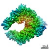
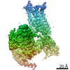
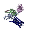
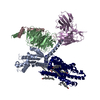
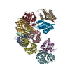
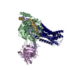
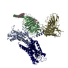
| Title | Crystal structure of mammalian Ric-8A:Galpha(i):nanobody complex | ||||||||||||
 Components Components |
| ||||||||||||
 Keywords Keywords |  SIGNALING PROTEIN / Ric-8A / G protein / GEF SIGNALING PROTEIN / Ric-8A / G protein / GEF | ||||||||||||
| Function / homology |  Function and homology information Function and homology informationcell-cell adhesion involved in gastrulation / cell migration involved in gastrulation / Extra-nuclear estrogen signaling / Adenylate cyclase inhibitory pathway / basement membrane organization / vasculature development / Adrenaline,noradrenaline inhibits insulin secretion / ADP signalling through P2Y purinoceptor 12 / G alpha (i) signalling events / negative regulation of synaptic transmission ...cell-cell adhesion involved in gastrulation / cell migration involved in gastrulation / Extra-nuclear estrogen signaling / Adenylate cyclase inhibitory pathway / basement membrane organization / vasculature development / Adrenaline,noradrenaline inhibits insulin secretion / ADP signalling through P2Y purinoceptor 12 / G alpha (i) signalling events / negative regulation of synaptic transmission / GTPase activating protein binding / positive regulation of protein localization to cell cortex / G-protein alpha-subunit binding / regulation of cAMP-mediated signaling / D2 dopamine receptor binding / G protein-coupled serotonin receptor binding / regulation of mitotic spindle organization / cellular response to forskolin / adenylate cyclase-inhibiting G protein-coupled receptor signaling pathway / guanyl-nucleotide exchange factor activity / G protein-coupled receptor binding / G-protein beta/gamma-subunit complex binding / visual learning / adenylate cyclase-modulating G protein-coupled receptor signaling pathway / GDP binding / heterotrimeric G-protein complex / cell cortex / midbody / in utero embryonic development / cell cycle / G protein-coupled receptor signaling pathway / cell division / GTPase activity / centrosome / GTP binding / magnesium ion binding / protein-containing complex / nucleus / plasma membrane / cytoplasmSimilarity search - Function | ||||||||||||
| Biological species |  Rattus norvegicus (Norway rat)Lama glama (llama) Rattus norvegicus (Norway rat)Lama glama (llama) | ||||||||||||
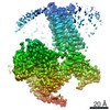
| Method | X-RAY DIFFRACTION / SYNCHROTRON / MOLECULAR REPLACEMENT / Resolution: 3.3 Å | ||||||||||||
 Authors Authors | Mou, T.C. / McClelland, L. / Yates-Hansen, C. / Sprang, S.R. | ||||||||||||
| Funding support |  United States, 3items United States, 3items
| ||||||||||||
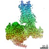
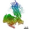
 Citation Citation | Journal: Nat Commun / Year: 2020 Title: Structure of the G protein chaperone and guanine nucleotide exchange factor Ric-8A bound to Gαi1. Authors: Levi J McClelland / Kaiming Zhang / Tung-Chung Mou / Jake Johnston / Cindee Yates-Hansen / Shanshan Li / Celestine J Thomas / Tzanko I Doukov / Sarah Triest / Alexandre Wohlkonig / Gregory G ...Authors: Levi J McClelland / Kaiming Zhang / Tung-Chung Mou / Jake Johnston / Cindee Yates-Hansen / Shanshan Li / Celestine J Thomas / Tzanko I Doukov / Sarah Triest / Alexandre Wohlkonig / Gregory G Tall / Jan Steyaert / Wah Chiu / Stephen R Sprang /  Abstract: Ric-8A is a cytosolic Guanine Nucleotide exchange Factor (GEF) that activates heterotrimeric G protein alpha subunits (Gα) and serves as an essential Gα chaperone. Mechanisms by which Ric-8A ...Ric-8A is a cytosolic Guanine Nucleotide exchange Factor (GEF) that activates heterotrimeric G protein alpha subunits (Gα) and serves as an essential Gα chaperone. Mechanisms by which Ric-8A catalyzes these activities, which are stimulated by Casein Kinase II phosphorylation, are unknown. We report the structure of the nanobody-stabilized complex of nucleotide-free Gα bound to phosphorylated Ric-8A at near atomic resolution by cryo-electron microscopy and X-ray crystallography. The mechanism of Ric-8A GEF activity differs considerably from that employed by G protein-coupled receptors at the plasma membrane. Ric-8A engages a specific conformation of Gα at multiple interfaces to form a complex that is stabilized by phosphorylation within a Ric-8A segment that connects two Gα binding sites. The C-terminus of Gα is ejected from its beta sheet core, thereby dismantling the GDP binding site. Ric-8A binds to the exposed Gα beta sheet and switch II to stabilize the nucleotide-free state of Gα. | ||||||||||||
| History |
|
- Structure visualization
| Structure viewer | Molecule: MolmilJmol/JSmol |
|---|
- Downloads & links
-Download
| PDBx/mmCIF format | 6tyl.cif.gz | 816.1 KB | Display | PDBx/mmCIF format |
|---|---|---|---|---|
| PDB format | pdb6tyl.ent.gz | 680.6 KB | Display | PDB format |
| PDBx/mmJSON format | 6tyl.json.gz | Tree view | PDBx/mmJSON format | |
| Others |  Other downloads Other downloads |
-Validation report
| Arichive directory | https://data.pdbj.org/pub/pdb/validation_reports/ty/6tylftp://data.pdbj.org/pub/pdb/validation_reports/ty/6tyl | HTTPS FTP |
|---|
-Related structure data
| Related structure data |  6uktC 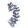 6nmgS S: Starting model for refinement C: citing same article ( |
|---|---|
| Similar structure data |

-Links
 PDBj
PDBj


- Assembly
| Deposited unit | 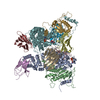
| ||||||||||
|---|---|---|---|---|---|---|---|---|---|---|---|
| 1 | 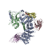
| ||||||||||
| 2 | 
| ||||||||||
| Unit cell |
|
-Components
| #1: Protein | Mass: 55836.926 Da / Num. of mol.: 2 / Mutation: Y232F Source method: isolated from a genetically manipulated source Source: (gene. exp.) Rattus norvegicus (Norway rat) / Gene: Ric8a, rCG_48458 / Production host:  Escherichia coli (E. coli) / References: UniProt: B1H241 Escherichia coli (E. coli) / References: UniProt: B1H241#2: Antibody | Mass: 13609.938 Da / Num. of mol.: 2 Source method: isolated from a genetically manipulated source Source: (gene. exp.) Lama glama (llama) / Production host: Escherichia coli (E. coli)#3: Antibody | Mass: 14247.603 Da / Num. of mol.: 2 Source method: isolated from a genetically manipulated source Source: (gene. exp.) Lama glama (llama) / Production host: Escherichia coli (E. coli)#4: Antibody | Mass: 14894.400 Da / Num. of mol.: 2 Source method: isolated from a genetically manipulated source Source: (gene. exp.) Lama glama (llama) / Production host: Escherichia coli (E. coli)#5: Protein | Mass: 40399.031 Da / Num. of mol.: 2 Source method: isolated from a genetically manipulated source Source: (gene. exp.) Rattus norvegicus (Norway rat) / Gene: Gnai1, Gnai-1 / Production host: Escherichia coli (E. coli) / References: UniProt: P10824Has ligand of interest | Y | |
|---|
-Experimental details
-Experiment
| Experiment | Method: X-RAY DIFFRACTION / Number of used crystals: 1 |
|---|
- Sample preparation
| Crystal | Density Matthews: 2.76 Å3/Da / Density % sol: 55.47 % |
|---|---|
| Crystal grow | Temperature: 285 K / Method: vapor diffusion, hanging drop / Details: 1.2-1.4M Sodium Malonate / PH range: 6-8 |
-Data collection
| Diffraction | Mean temperature: 100 K / Serial crystal experiment: N |
|---|---|
| Diffraction source | Source: SYNCHROTRON / Site: NSLS-II / Beamline: 17-ID-2 / Wavelength: 0.987 Å |
| Detector | Type: DECTRIS EIGER X 16M / Detector: PIXEL / Date: Nov 9, 2018 |
| Radiation | Monochromator: Si(111) / Protocol: SINGLE WAVELENGTH / Monochromatic (M) / Laue (L): M / Scattering type: x-ray |
| Radiation wavelength | Wavelength: 0.987 Å / Relative weight: 1 |
| Reflection | Resolution: 3.3→39.58 Å / Num. obs: 20707 / % possible obs: 90.1 % / Redundancy: 3.6 % / Biso Wilson estimate: 91.88 Å2 / CC1/2: 0.99 / Rpim(I) all: 0.11 / Rrim(I) all: 0.21 / Net I/σ(I): 3.7 |
| Reflection shell | Resolution: 3.3→3.9 Å / Mean I/σ(I) obs: 1.7 / Num. unique obs: 1035 / CC1/2: 0.7 / Rpim(I) all: 0.38 / Rrim(I) all: 0.7 |
- Processing
| Software |
| ||||||||||||||||||||||||||||||||||||||||
|---|---|---|---|---|---|---|---|---|---|---|---|---|---|---|---|---|---|---|---|---|---|---|---|---|---|---|---|---|---|---|---|---|---|---|---|---|---|---|---|---|---|
| Refinement | Method to determine structure: MOLECULAR REPLACEMENT Starting model: 6NMG Resolution: 3.3→39.53 Å / Cross valid method: FREE R-VALUE
| ||||||||||||||||||||||||||||||||||||||||
| Displacement parameters | Biso mean: 109.9 Å2 | ||||||||||||||||||||||||||||||||||||||||
| Refinement step | Cycle: LAST / Resolution: 3.3→39.53 Å
| ||||||||||||||||||||||||||||||||||||||||
| Refine LS restraints |
| ||||||||||||||||||||||||||||||||||||||||
| Refinement TLS params. | Method: refined / Origin x: 25.8389 Å / Origin y: -18.0538 Å / Origin z: 27.0874 Å
| ||||||||||||||||||||||||||||||||||||||||
| Refinement TLS group | Selection details: all |
