 Movie
Movie Controller

[English] 日本語
 Yorodumi
- PDB-6twc: Crystal Structure of the Catalytic Domain of the Coagulation Fact... -
+ Open data

- Basic information
| Entry | Database: PDB / ID: 6twc | ||||||
|---|---|---|---|---|---|---|---|
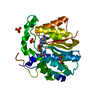
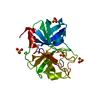
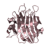
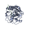
| Title | Crystal Structure of the Catalytic Domain of the Coagulation Factor XIa in Complex with Double Bridged Peptide F21 | ||||||
 Components Components |
| ||||||
 Keywords Keywords |  BLOOD CLOTTING / Protease / Coagulation factors / Inhibitor / Double bridged peptide / Phage display BLOOD CLOTTING / Protease / Coagulation factors / Inhibitor / Double bridged peptide / Phage display | ||||||
| Function / homology |  Function and homology informationcoagulation factor XIa / serine-type aminopeptidase activity / Defective F9 activation / positive regulation of fibrinolysis / plasminogen activation / Intrinsic Pathway of Fibrin Clot Formation / blood coagulation / heparin binding / serine-type endopeptidase activity / extracellular space ...coagulation factor XIa / serine-type aminopeptidase activity / Defective F9 activation / positive regulation of fibrinolysis / plasminogen activation / Intrinsic Pathway of Fibrin Clot Formation / blood coagulation / heparin binding / serine-type endopeptidase activity / extracellular space / extracellular exosome / extracellular region / membrane / identical protein binding / plasma membrane Function and homology informationcoagulation factor XIa / serine-type aminopeptidase activity / Defective F9 activation / positive regulation of fibrinolysis / plasminogen activation / Intrinsic Pathway of Fibrin Clot Formation / blood coagulation / heparin binding / serine-type endopeptidase activity / extracellular space ...coagulation factor XIa / serine-type aminopeptidase activity / Defective F9 activation / positive regulation of fibrinolysis / plasminogen activation / Intrinsic Pathway of Fibrin Clot Formation / blood coagulation / heparin binding / serine-type endopeptidase activity / extracellular space / extracellular exosome / extracellular region / membrane / identical protein binding / plasma membraneSimilarity search - Function | ||||||
| Biological species |  Homo sapiens (human) Homo sapiens (human)synthetic construct (others) | ||||||
| Method | X-RAY DIFFRACTION / SYNCHROTRON / MOLECULAR REPLACEMENT / Resolution: 2.86 Å | ||||||
 Authors Authors | Kong, X.D. / Pojer, F. / Heinis, C. | ||||||
| Funding support |  Switzerland, 1items Switzerland, 1items
| ||||||
 Citation Citation | Journal: Nat Biomed Eng / Year: 2020 Title: De novo development of proteolytically resistant therapeutic peptides for oral administration. Authors: Kong, X.D. / Moriya, J. / Carle, V. / Pojer, F. / Abriata, L.A. / Deyle, K. / Heinis, C. | ||||||
| History |
|
- Structure visualization
| Structure viewer | Molecule: MolmilJmol/JSmol |
|---|
- Downloads & links
-Download
| PDBx/mmCIF format | 6twc.cif.gz | 67.4 KB | Display | PDBx/mmCIF format |
|---|---|---|---|---|
| PDB format | pdb6twc.ent.gz | 47.8 KB | Display | PDB format |
| PDBx/mmJSON format | 6twc.json.gz | Tree view | PDBx/mmJSON format | |
| Others |  Other downloads Other downloads |
-Validation report
| Arichive directory | https://data.pdbj.org/pub/pdb/validation_reports/tw/6twcftp://data.pdbj.org/pub/pdb/validation_reports/tw/6twc | HTTPS FTP |
|---|
-Related structure data
| Related structure data |  6twbC  4ty6S S: Starting model for refinement C: citing same article ( |
|---|---|
| Similar structure data |


-Links
 PDBj
PDBj


- Assembly
| Deposited unit | 
| ||||||||
|---|---|---|---|---|---|---|---|---|---|
| 1 |
| ||||||||
| Unit cell |
|
-Components
| #1: Protein | Factor XI / FXI / Plasma thromboplastin antecedent / PTA Mass: 27600.342 Da / Num. of mol.: 1 Source method: isolated from a genetically manipulated source Source: (gene. exp.) Homo sapiens (human) / Gene: F11 / Production host:  Escherichia coli (E. coli) / References: UniProt: P03951, coagulation factor XIa Escherichia coli (E. coli) / References: UniProt: P03951, coagulation factor XIa | ||
|---|---|---|---|
| #2: Protein/peptide | Factor XI / FXI / Plasma thromboplastin antecedent / PTA Mass: 2159.419 Da / Num. of mol.: 1 Source method: isolated from a genetically manipulated source Source: (gene. exp.) Homo sapiens (human) / Gene: F11 / Production host: Escherichia coli (E. coli) / References: UniProt: P03951, coagulation factor XIa | ||
| #3: Protein/peptide | Mass: 1428.811 Da / Num. of mol.: 1 / Source method: obtained synthetically / Source: (synth.) synthetic construct (others) | ||
| #4: Chemical | Acetone  Mass: 58.079 Da / Num. of mol.: 2 / Source method: obtained synthetically / Formula: C3H6O / Feature type: SUBJECT OF INVESTIGATION Mass: 58.079 Da / Num. of mol.: 2 / Source method: obtained synthetically / Formula: C3H6O / Feature type: SUBJECT OF INVESTIGATIONHas ligand of interest | Y | |
-Experimental details
-Experiment
| Experiment | Method: X-RAY DIFFRACTION / Number of used crystals: 1 |
|---|
- Sample preparation
| Crystal | Density Matthews: 3.14 Å3/Da / Density % sol: 60.86 % |
|---|---|
| Crystal grow | Temperature: 291 K / Method: vapor diffusion, hanging drop Details: 200 mM (NH4)2SO4, 25% PEG4000, and 100 mM NaOAc, pH 4.6 |
-Data collection
| Diffraction | Mean temperature: 100 K / Serial crystal experiment: N | ||||||||||||||||||||||||||||||||||||||||||||||||||||||||||||||||||||||||||||||||||||||||||||||||||||
|---|---|---|---|---|---|---|---|---|---|---|---|---|---|---|---|---|---|---|---|---|---|---|---|---|---|---|---|---|---|---|---|---|---|---|---|---|---|---|---|---|---|---|---|---|---|---|---|---|---|---|---|---|---|---|---|---|---|---|---|---|---|---|---|---|---|---|---|---|---|---|---|---|---|---|---|---|---|---|---|---|---|---|---|---|---|---|---|---|---|---|---|---|---|---|---|---|---|---|---|---|---|
| Diffraction source | Source: SYNCHROTRON / Site: SLS / Beamline: X06DA / Wavelength: 1 Å | ||||||||||||||||||||||||||||||||||||||||||||||||||||||||||||||||||||||||||||||||||||||||||||||||||||
| Detector | Type: DECTRIS PILATUS 2M-F / Detector: PIXEL / Date: Dec 20, 2017 | ||||||||||||||||||||||||||||||||||||||||||||||||||||||||||||||||||||||||||||||||||||||||||||||||||||
| Radiation | Protocol: SINGLE WAVELENGTH / Monochromatic (M) / Laue (L): M / Scattering type: x-ray | ||||||||||||||||||||||||||||||||||||||||||||||||||||||||||||||||||||||||||||||||||||||||||||||||||||
| Radiation wavelength | Wavelength: 1 Å / Relative weight: 1 | ||||||||||||||||||||||||||||||||||||||||||||||||||||||||||||||||||||||||||||||||||||||||||||||||||||
| Reflection | Resolution: 2.86→43.783 Å / Num. obs: 9502 / % possible obs: 99.6 % / Redundancy: 12.301 % / Biso Wilson estimate: 56.14 Å2 / CC1/2: 0.999 / Rmerge(I) obs: 0.124 / Rrim(I) all: 0.13 / Χ2: 1.053 / Net I/σ(I): 22.33 / Num. measured all: 116888 | ||||||||||||||||||||||||||||||||||||||||||||||||||||||||||||||||||||||||||||||||||||||||||||||||||||
| Reflection shell | Diffraction-ID: 1
|
- Processing
| Software |
| ||||||||||||||||||||||||||||||
|---|---|---|---|---|---|---|---|---|---|---|---|---|---|---|---|---|---|---|---|---|---|---|---|---|---|---|---|---|---|---|---|
| Refinement | Method to determine structure: MOLECULAR REPLACEMENT Starting model: 4TY6 Resolution: 2.86→43.782 Å / SU ML: 0.37 / Cross valid method: THROUGHOUT / σ(F): 1.34 / Phase error: 28.99 / Stereochemistry target values: ML
| ||||||||||||||||||||||||||||||
| Solvent computation | Shrinkage radii: 0.9 Å / VDW probe radii: 1.11 Å / Solvent model: FLAT BULK SOLVENT MODEL | ||||||||||||||||||||||||||||||
| Displacement parameters | Biso max: 132.95 Å2 / Biso mean: 67.8712 Å2 / Biso min: 43.23 Å2 | ||||||||||||||||||||||||||||||
| Refinement step | Cycle: final / Resolution: 2.86→43.782 Å
| ||||||||||||||||||||||||||||||
| LS refinement shell | Refine-ID: X-RAY DIFFRACTION / Rfactor Rfree error: 0
|
