 Movie
Movie Controller

[English] 日本語
 Yorodumi
- PDB-6tqu: The crystal structure of the MSP domain of human MOSPD2 in comple... -
+ Open data

- Basic information
| Entry | Database: PDB / ID: 6tqu | |||||||||||||||
|---|---|---|---|---|---|---|---|---|---|---|---|---|---|---|---|---|
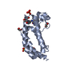
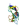
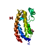
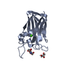
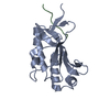
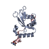
| Title | The crystal structure of the MSP domain of human MOSPD2 in complex with the Phospho-FFAT motif of STARD3. | |||||||||||||||
 Components Components |
| |||||||||||||||
 Keywords Keywords |  PROTEIN BINDING / Membrane contact sites / FFAT motif / MSP domain / Endoplasmic reticulum PROTEIN BINDING / Membrane contact sites / FFAT motif / MSP domain / Endoplasmic reticulum | |||||||||||||||
| Function / homology |  Function and homology information Function and homology informationvesicle tethering to endoplasmic reticulum / endoplasmic reticulum-endosome membrane contact site / progesterone biosynthetic process / FFAT motif binding / lipid droplet formation / organelle membrane contact site / Pregnenolone biosynthesis / cholesterol transfer activity / cholesterol transport / RHOD GTPase cycle ...vesicle tethering to endoplasmic reticulum / endoplasmic reticulum-endosome membrane contact site / progesterone biosynthetic process / FFAT motif binding / lipid droplet formation / organelle membrane contact site / Pregnenolone biosynthesis / cholesterol transfer activity / cholesterol transport / RHOD GTPase cycle / positive regulation of neutrophil chemotaxis / positive regulation of monocyte chemotaxis / cholesterol binding / mitochondrial transport / steroid metabolic process / endomembrane system / specific granule membrane / cholesterol metabolic process / protein homooligomerization / lipid metabolic process / chemotaxis / late endosome membrane / endosome / lysosomal membrane / intracellular membrane-bounded organelle / Neutrophil degranulation / endoplasmic reticulum membrane / endoplasmic reticulum / protein homodimerization activity / mitochondrion / nucleoplasm / membrane / plasma membrane / cytosol / cytoplasmSimilarity search - Function | |||||||||||||||
| Biological species |  Homo sapiens (human) Homo sapiens (human) | |||||||||||||||
| Method | X-RAY DIFFRACTION / SYNCHROTRON / MOLECULAR REPLACEMENT / molecular replacement / Resolution: 2.4 Å | |||||||||||||||
 Authors Authors | McEwen, A.G. / Poussin-Courmontagne, P. / Di Mattia, T. / Wendling, C. / Cavarelli, J. / Tomasetto, C. / Alpy, F. | |||||||||||||||
| Funding support |  France, 4items France, 4items
| |||||||||||||||
 Citation Citation | Journal: Embo J. / Year: 2020 Title: FFAT motif phosphorylation controls formation and lipid transfer function of inter-organelle contacts. Authors: Di Mattia, T. / Martinet, A. / Ikhlef, S. / McEwen, A.G. / Nomine, Y. / Wendling, C. / Poussin-Courmontagne, P. / Voilquin, L. / Eberling, P. / Ruffenach, F. / Cavarelli, J. / Slee, J. / ...Authors: Di Mattia, T. / Martinet, A. / Ikhlef, S. / McEwen, A.G. / Nomine, Y. / Wendling, C. / Poussin-Courmontagne, P. / Voilquin, L. / Eberling, P. / Ruffenach, F. / Cavarelli, J. / Slee, J. / Levine, T.P. / Drin, G. / Tomasetto, C. / Alpy, F. | |||||||||||||||
| History |
|
- Structure visualization
| Structure viewer | Molecule: MolmilJmol/JSmol |
|---|
- Downloads & links
-Download
| PDBx/mmCIF format | 6tqu.cif.gz | 125.8 KB | Display | PDBx/mmCIF format |
|---|---|---|---|---|
| PDB format | pdb6tqu.ent.gz | 97.8 KB | Display | PDB format |
| PDBx/mmJSON format | 6tqu.json.gz | Tree view | PDBx/mmJSON format | |
| Others |  Other downloads Other downloads |
-Validation report
| Arichive directory | https://data.pdbj.org/pub/pdb/validation_reports/tq/6tquftp://data.pdbj.org/pub/pdb/validation_reports/tq/6tqu | HTTPS FTP |
|---|
-Related structure data
| Related structure data | 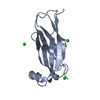 6tqrC  6tqsC  6tqtSC S: Starting model for refinement C: citing same article ( |
|---|---|
| Similar structure data |


-Links
 PDBj
PDBj- Assembly
| Deposited unit | 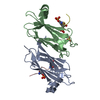
| ||||||||
|---|---|---|---|---|---|---|---|---|---|
| 1 | 
| ||||||||
| 2 | 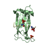
| ||||||||
| Unit cell |
| ||||||||
| Components on special symmetry positions |
|
-Components
| #1: Protein | Mass: 14833.074 Da / Num. of mol.: 2 Source method: isolated from a genetically manipulated source Details: MSP domain / Source: (gene. exp.) Homo sapiens (human) / Gene: MOSPD2 / Production host:  Escherichia coli BL21(DE3) (bacteria) / References: UniProt: Q8NHP6 Escherichia coli BL21(DE3) (bacteria) / References: UniProt: Q8NHP6#2: Protein/peptide | Mass: 2434.314 Da / Num. of mol.: 2 / Source method: obtained synthetically / Details: phospho-FFAT motif / Source: (synth.) Homo sapiens (human) / References: UniProt: Q14849#3: Chemical | ChemComp-SO4 / | Sulfate  Mass: 96.063 Da / Num. of mol.: 1 / Source method: obtained synthetically / Formula: SO4 Mass: 96.063 Da / Num. of mol.: 1 / Source method: obtained synthetically / Formula: SO4#4: Water | ChemComp-HOH / | Water Mass: 18.015 Da / Num. of mol.: 89 / Source method: isolated from a natural source / Formula: H2O Mass: 18.015 Da / Num. of mol.: 89 / Source method: isolated from a natural source / Formula: H2OHas ligand of interest | N | |
|---|
-Experimental details
-Experiment
| Experiment | Method: X-RAY DIFFRACTION / Number of used crystals: 1 |
|---|
- Sample preparation
| Crystal | Density Matthews: 1.97 Å3/Da / Density % sol: 37.5 % |
|---|---|
| Crystal grow | Temperature: 293 K / Method: vapor diffusion, sitting drop / pH: 7.5 Details: 7.5% PEG 2000, 7.5% PEG 3350, 7.5% PEG 4000, 7.5% PEG 5000 MME, 0.05 M Magnesium sulfate, 0.1 M HEPES pH 7.5 |
-Data collection
| Diffraction | Mean temperature: 100 K / Serial crystal experiment: N | ||||||||||||||||||||||||||||||
|---|---|---|---|---|---|---|---|---|---|---|---|---|---|---|---|---|---|---|---|---|---|---|---|---|---|---|---|---|---|---|---|
| Diffraction source | Source: SYNCHROTRON / Site: SOLEIL / Beamline: PROXIMA 2 / Wavelength: 0.9801 Å | ||||||||||||||||||||||||||||||
| Detector | Type: DECTRIS EIGER X 9M / Detector: PIXEL / Date: Nov 24, 2018 | ||||||||||||||||||||||||||||||
| Radiation | Monochromator: Si(111) / Protocol: SINGLE WAVELENGTH / Monochromatic (M) / Laue (L): M / Scattering type: x-ray | ||||||||||||||||||||||||||||||
| Radiation wavelength | Wavelength: 0.9801 Å / Relative weight: 1 | ||||||||||||||||||||||||||||||
| Reflection | Resolution: 2.4→87.97 Å / Num. obs: 15203 / % possible obs: 100 % / Redundancy: 24.9 % / CC1/2: 0.997 / Rmerge(I) obs: 0.258 / Rpim(I) all: 0.053 / Rrim(I) all: 0.263 / Net I/σ(I): 9.1 / Num. measured all: 378156 / Scaling rejects: 6 | ||||||||||||||||||||||||||||||
| Reflection shell | Diffraction-ID: 1
|
-Phasing
| Phasing | Method: molecular replacement | |||||||||
|---|---|---|---|---|---|---|---|---|---|---|
| Phasing MR |
|
- Processing
| Software |
| |||||||||||||||||||||||||||||||||||||||||||||||||||||||||||||||||||||||||||||||||||||||||||||||||||||||||||||||||||||||||||||
|---|---|---|---|---|---|---|---|---|---|---|---|---|---|---|---|---|---|---|---|---|---|---|---|---|---|---|---|---|---|---|---|---|---|---|---|---|---|---|---|---|---|---|---|---|---|---|---|---|---|---|---|---|---|---|---|---|---|---|---|---|---|---|---|---|---|---|---|---|---|---|---|---|---|---|---|---|---|---|---|---|---|---|---|---|---|---|---|---|---|---|---|---|---|---|---|---|---|---|---|---|---|---|---|---|---|---|---|---|---|---|---|---|---|---|---|---|---|---|---|---|---|---|---|---|---|---|
| Refinement | Method to determine structure: MOLECULAR REPLACEMENT Starting model: 6TQT Resolution: 2.4→43.98 Å / SU ML: 0.3 / Cross valid method: THROUGHOUT / σ(F): 1.36 / Phase error: 27.86
| |||||||||||||||||||||||||||||||||||||||||||||||||||||||||||||||||||||||||||||||||||||||||||||||||||||||||||||||||||||||||||||
| Solvent computation | Shrinkage radii: 0.9 Å / VDW probe radii: 1.11 Å | |||||||||||||||||||||||||||||||||||||||||||||||||||||||||||||||||||||||||||||||||||||||||||||||||||||||||||||||||||||||||||||
| Displacement parameters | Biso max: 180.95 Å2 / Biso mean: 75.1558 Å2 / Biso min: 36.87 Å2 | |||||||||||||||||||||||||||||||||||||||||||||||||||||||||||||||||||||||||||||||||||||||||||||||||||||||||||||||||||||||||||||
| Refinement step | Cycle: final / Resolution: 2.4→43.98 Å
| |||||||||||||||||||||||||||||||||||||||||||||||||||||||||||||||||||||||||||||||||||||||||||||||||||||||||||||||||||||||||||||
| LS refinement shell | Refine-ID: X-RAY DIFFRACTION / Rfactor Rfree error: 0 / Total num. of bins used: 5 / % reflection obs: 100 %
| |||||||||||||||||||||||||||||||||||||||||||||||||||||||||||||||||||||||||||||||||||||||||||||||||||||||||||||||||||||||||||||
| Refinement TLS params. | Method: refined / Refine-ID: X-RAY DIFFRACTION
| |||||||||||||||||||||||||||||||||||||||||||||||||||||||||||||||||||||||||||||||||||||||||||||||||||||||||||||||||||||||||||||
| Refinement TLS group |
|
