 Movie
Movie Controller


+ Open data

- Basic information
| Entry | Database: PDB / ID: 6tpi | |||||||||
|---|---|---|---|---|---|---|---|---|---|---|
| Title | EnvC bound to the FtsX periplasmic domain | |||||||||
 Components Components |
| |||||||||
 Keywords Keywords |  PROTEIN BINDING / Complex PROTEIN BINDING / Complex | |||||||||
| Function / homology |  Function and homology information Function and homology informationdivision septum / divisome complex / peptidoglycan-based cell wall biogenesis / Gram-negative-bacterium-type cell wall / septum digestion after cytokinesis / peptidoglycan turnover / plasma membrane protein complex / FtsZ-dependent cytokinesis / division septum assembly / cell division site ...division septum / divisome complex / peptidoglycan-based cell wall biogenesis / Gram-negative-bacterium-type cell wall / septum digestion after cytokinesis / peptidoglycan turnover / plasma membrane protein complex / FtsZ-dependent cytokinesis / division septum assembly / cell division site / ATPase complex / positive regulation of cell division / response to radiation / metalloendopeptidase activity / outer membrane-bounded periplasmic space / periplasmic space / hydrolase activity / response to xenobiotic stimulus / cell cycle / cell division / plasma membraneSimilarity search - Function | |||||||||
| Biological species |  Escherichia coli (E. coli) Escherichia coli (E. coli) | |||||||||
| Method | X-RAY DIFFRACTION / SYNCHROTRON / MOLECULAR REPLACEMENT / molecular replacement / Resolution: 2.1 Å | |||||||||
 Authors Authors | Crow, A. | |||||||||
| Funding support |  United Kingdom, 2items United Kingdom, 2items
| |||||||||
 Citation Citation | Journal: Proc.Natl.Acad.Sci.USA / Year: 2020 Title: Insights into bacterial cell division from a structure of EnvC bound to the FtsX periplasmic domain. Authors: Cook, J. / Baverstock, T.C. / McAndrew, M.B.L. / Stansfeld, P.J. / Roper, D.I. / Crow, A. | |||||||||
| History |
|
- Structure visualization
| Structure viewer | Molecule: MolmilJmol/JSmol |
|---|
- Downloads & links
-Download
| PDBx/mmCIF format | 6tpi.cif.gz | 132.6 KB | Display | PDBx/mmCIF format |
|---|---|---|---|---|
| PDB format | pdb6tpi.ent.gz | 101 KB | Display | PDB format |
| PDBx/mmJSON format | 6tpi.json.gz | Tree view | PDBx/mmJSON format | |
| Others |  Other downloads Other downloads |
-Validation report
| Arichive directory | https://data.pdbj.org/pub/pdb/validation_reports/tp/6tpiftp://data.pdbj.org/pub/pdb/validation_reports/tp/6tpi | HTTPS FTP |
|---|
-Related structure data
| Related structure data | 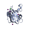 4bh5S  4n8oS S: Starting model for refinement |
|---|---|
| Similar structure data | |
| Experimental dataset #1 | Data reference: 10.5281/zenodo.3574434 / Data set type: diffraction image data |
-Links
 PDBj
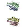
PDBj- Assembly
| Deposited unit | 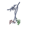
| ||||||||||||||||||
|---|---|---|---|---|---|---|---|---|---|---|---|---|---|---|---|---|---|---|---|
| 1 |
| ||||||||||||||||||
| Unit cell |
| ||||||||||||||||||
| Noncrystallographic symmetry (NCS) | NCS domain:
NCS domain segments: Component-ID: 0 / Ens-ID: 1 / Beg auth comp-ID: GLN / Beg label comp-ID: GLN / End auth comp-ID: ARG / End label comp-ID: ARG / Refine code: 0 / Auth seq-ID: 110 - 205 / Label seq-ID: 3 - 98
|
-Components
| #1: Protein | Mass: 43117.344 Da / Num. of mol.: 1 Source method: isolated from a genetically manipulated source Source: (gene. exp.) Escherichia coli (strain K12) (bacteria)Gene: envC, yibP, b3613, JW5646 / Production host: Escherichia coli BL21(DE3) (bacteria) / Variant (production host): C43 / References: UniProt: P37690 | ||
|---|---|---|---|
| #2: Protein | Mass: 12262.586 Da / Num. of mol.: 2 Source method: isolated from a genetically manipulated source Source: (gene. exp.) Escherichia coli (strain K12) (bacteria)Gene: ftsX, ftsS, b3462, JW3427 / Production host: Escherichia coli BL21(DE3) (bacteria) / Variant (production host): C43 / References: UniProt: P0AC30#3: Water | ChemComp-HOH / | Water Mass: 18.015 Da / Num. of mol.: 229 / Source method: isolated from a natural source / Formula: H2O Mass: 18.015 Da / Num. of mol.: 229 / Source method: isolated from a natural source / Formula: H2O |
-Experimental details
-Experiment
| Experiment | Method: X-RAY DIFFRACTION / Number of used crystals: 1 |
|---|
- Sample preparation
| Crystal | Density Matthews: 2.68 Å3/Da / Density % sol: 54.15 % |
|---|---|
| Crystal grow | Temperature: 298 K / Method: vapor diffusion, sitting drop / pH: 8.5 Details: 10% PEG 8000, 20 % Ethylene glycol, 0.1 M Tris/bicine pH8.5, ) 0.12M monosaccarides (glucose, mannose, galactose, fucose, xylose, NAG) |
-Data collection
| Diffraction | Mean temperature: 100 K / Serial crystal experiment: N |
|---|---|
| Diffraction source | Source: SYNCHROTRON / Site: Diamond / Beamline: I04-1 / Wavelength: 0.91587 Å |
| Detector | Type: DECTRIS PILATUS 6M-F / Detector: PIXEL / Date: Apr 7, 2019 |
| Radiation | Protocol: SINGLE WAVELENGTH / Monochromatic (M) / Laue (L): M / Scattering type: x-ray |
| Radiation wavelength | Wavelength: 0.91587 Å / Relative weight: 1 |
| Reflection | Resolution: 2.1→55.823 Å / Num. obs: 43252 / % possible obs: 100 % / Redundancy: 12 % / CC1/2: 0.999 / Rpim(I) all: 0.044 / Net I/σ(I): 15 |
| Reflection shell | Resolution: 2.1→2.16 Å / Redundancy: 10.8 % / Mean I/σ(I) obs: 2.2 / Num. unique obs: 3494 / CC1/2: 0.767 / Rpim(I) all: 0.577 / % possible all: 100 |
-Phasing
| Phasing | Method: molecular replacement |
|---|
- Processing
| Software |
| ||||||||||||||||||||||||||||||||||||||||||||||||||||||||||||
|---|---|---|---|---|---|---|---|---|---|---|---|---|---|---|---|---|---|---|---|---|---|---|---|---|---|---|---|---|---|---|---|---|---|---|---|---|---|---|---|---|---|---|---|---|---|---|---|---|---|---|---|---|---|---|---|---|---|---|---|---|---|
| Refinement | Method to determine structure: MOLECULAR REPLACEMENT Starting model: 4BH5 and 4N8O Resolution: 2.1→55.82 Å / Cor.coef. Fo:Fc: 0.954 / Cor.coef. Fo:Fc free: 0.921 / SU B: 7.669 / SU ML: 0.187 / Cross valid method: THROUGHOUT / σ(F): 0 / ESU R: 0.213 / ESU R Free: 0.2 Details: HYDROGENS HAVE BEEN ADDED IN THE RIDING POSITIONS U VALUES : REFINED INDIVIDUALLY
| ||||||||||||||||||||||||||||||||||||||||||||||||||||||||||||
| Solvent computation | Ion probe radii: 0.8 Å / Shrinkage radii: 0.8 Å / VDW probe radii: 1.2 Å | ||||||||||||||||||||||||||||||||||||||||||||||||||||||||||||
| Displacement parameters | Biso max: 209.69 Å2 / Biso mean: 58.531 Å2 / Biso min: 25.33 Å2
| ||||||||||||||||||||||||||||||||||||||||||||||||||||||||||||
| Refinement step | Cycle: final / Resolution: 2.1→55.82 Å
| ||||||||||||||||||||||||||||||||||||||||||||||||||||||||||||
| Refine LS restraints |
| ||||||||||||||||||||||||||||||||||||||||||||||||||||||||||||
| Refine LS restraints NCS | Ens-ID: 1 / Number: 2391 / Refine-ID: X-RAY DIFFRACTION / Type: interatomic distance / Rms dev position: 0.2 Å / Weight position: 0.05
| ||||||||||||||||||||||||||||||||||||||||||||||||||||||||||||
| LS refinement shell | Resolution: 2.1→2.155 Å / Rfactor Rfree error: 0 / Total num. of bins used: 20
|
