 Movie
Movie Controller


+ Open data

- Basic information
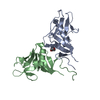
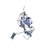
| Entry | Database: PDB / ID: 6tla | ||||||
|---|---|---|---|---|---|---|---|
| Title | CRYSTAL STRUCTURE OF LECTIN-LIKE OX-LDL RECEPTOR 1 (C 1 2 1) | ||||||
 Components Components | Oxidized low-density lipoprotein receptor 1 | ||||||
 Keywords Keywords | LIPID BINDING PROTEIN /  LOX-1 / OLR1 / CTLD / C-TYPE LECTIN LIKE DOMAIN / SCAVENGER RECEPTOR / OXLDL BINDING LOX-1 / OLR1 / CTLD / C-TYPE LECTIN LIKE DOMAIN / SCAVENGER RECEPTOR / OXLDL BINDING | ||||||
| Function / homology |  Function and homology information Function and homology informationlow-density lipoprotein particle receptor activity / lipoprotein metabolic process / blood circulation / leukocyte cell-cell adhesion / immune system process / tertiary granule membrane / specific granule membrane / Cell surface interactions at the vascular wall / carbohydrate binding / receptor complex ...low-density lipoprotein particle receptor activity / lipoprotein metabolic process / blood circulation / leukocyte cell-cell adhesion / immune system process / tertiary granule membrane / specific granule membrane / Cell surface interactions at the vascular wall / carbohydrate binding / receptor complex / inflammatory response / membrane raft / intracellular membrane-bounded organelle / Neutrophil degranulation / proteolysis / extracellular region / nucleoplasm / membrane / identical protein binding / plasma membraneSimilarity search - Function | ||||||
| Biological species |  Homo sapiens (human) Homo sapiens (human) | ||||||
| Method | X-RAY DIFFRACTION / SYNCHROTRON / MOLECULAR REPLACEMENT / Resolution: 2.16 Å | ||||||
 Authors Authors | Nar, H. / Fiegen, D. / Schnapp, G. | ||||||
 Citation Citation | Journal: Commun Chem / Year: 2020 Title: A small-molecule inhibitor of lectin-like oxidized LDL receptor-1 acts by stabilizing an inactive receptor tetramer state Authors: Nar, H. / Fiegen, D. / Schnapp, G. | ||||||
| History |
|
- Structure visualization
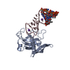
| Structure viewer | Molecule: MolmilJmol/JSmol |
|---|
- Downloads & links
-Download
| PDBx/mmCIF format | 6tla.cif.gz | 174.6 KB | Display | PDBx/mmCIF format |
|---|---|---|---|---|
| PDB format | pdb6tla.ent.gz | 140.4 KB | Display | PDB format |
| PDBx/mmJSON format | 6tla.json.gz | Tree view | PDBx/mmJSON format | |
| Others |  Other downloads Other downloads |
-Validation report
| Arichive directory | https://data.pdbj.org/pub/pdb/validation_reports/tl/6tlaftp://data.pdbj.org/pub/pdb/validation_reports/tl/6tla | HTTPS FTP |
|---|
-Related structure data
| Related structure data |  6tl7C  6tl9C  1ypuS S: Starting model for refinement C: citing same article ( |
|---|---|
| Similar structure data |


-Links
 PDBj
PDBj


- Assembly
| Deposited unit | 
| ||||||||
|---|---|---|---|---|---|---|---|---|---|
| 1 | 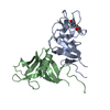
| ||||||||
| 2 | 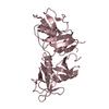
| ||||||||
| Unit cell |
|
-Components
| #1: Protein | Mass: 16910.203 Da / Num. of mol.: 3 Source method: isolated from a genetically manipulated source Source: (gene. exp.) Homo sapiens (human) / Gene: OLR1, CLEC8A, LOX1 / Production host:  Escherichia coli (E. coli) / References: UniProt: P78380 Escherichia coli (E. coli) / References: UniProt: P78380#2: Chemical | ChemComp-MES / | MES (buffer)  Mass: 195.237 Da / Num. of mol.: 1 / Source method: obtained synthetically / Formula: C6H13NO4S / Comment: pH buffer*YM Mass: 195.237 Da / Num. of mol.: 1 / Source method: obtained synthetically / Formula: C6H13NO4S / Comment: pH buffer*YM#3: Water | ChemComp-HOH / | Water Mass: 18.015 Da / Num. of mol.: 118 / Source method: isolated from a natural source / Formula: H2O Mass: 18.015 Da / Num. of mol.: 118 / Source method: isolated from a natural source / Formula: H2OHas ligand of interest | N | |
|---|
-Experimental details
-Experiment
| Experiment | Method: X-RAY DIFFRACTION / Number of used crystals: 1 |
|---|
- Sample preparation
| Crystal | Density Matthews: 2.45 Å3/Da / Density % sol: 49.89 % |
|---|---|
| Crystal grow | Temperature: 277 K / Method: vapor diffusion, hanging drop / Details: 0.1 M MES pH 6, 20% PEG6000, 0.2 M LiCl |
-Data collection
| Diffraction | Mean temperature: 100 K / Serial crystal experiment: N |
|---|---|
| Diffraction source | Source: SYNCHROTRON / Site: SLS  / Beamline: X06SA / Wavelength: 0.9101 Å / Beamline: X06SA / Wavelength: 0.9101 Å |
| Detector | Type: DECTRIS PILATUS3 6M / Detector: PIXEL / Date: Dec 3, 2009 |
| Radiation | Protocol: SINGLE WAVELENGTH / Monochromatic (M) / Laue (L): M / Scattering type: x-ray |
| Radiation wavelength | Wavelength: 0.9101 Å / Relative weight: 1 |
| Reflection | Resolution: 2.16→109.75 Å / Num. obs: 22997 / % possible obs: 97 % / Redundancy: 3.1 % / Rmerge(I) obs: 0.029 / Rpim(I) all: 0.035 / Rrim(I) all: 0.035 / Net I/σ(I): 19.2 |
| Reflection shell | Resolution: 2.16→2.27 Å / Redundancy: 2.5 % / Rmerge(I) obs: 0.446 / Mean I/σ(I) obs: 2 / Num. unique obs: 3126 / Rpim(I) all: 0.32 / Rrim(I) all: 0.552 / % possible all: 90.5 |
- Processing
| Software |
| ||||||||||||||||||||||||||||||||||||||||||||||||||||||||||||||||||||||||||||||||||||||||||||||||||||
|---|---|---|---|---|---|---|---|---|---|---|---|---|---|---|---|---|---|---|---|---|---|---|---|---|---|---|---|---|---|---|---|---|---|---|---|---|---|---|---|---|---|---|---|---|---|---|---|---|---|---|---|---|---|---|---|---|---|---|---|---|---|---|---|---|---|---|---|---|---|---|---|---|---|---|---|---|---|---|---|---|---|---|---|---|---|---|---|---|---|---|---|---|---|---|---|---|---|---|---|---|---|
| Refinement | Method to determine structure: MOLECULAR REPLACEMENT Starting model: 1ypu Resolution: 2.16→55 Å / Cor.coef. Fo:Fc: 0.944 / Cor.coef. Fo:Fc free: 0.936 / SU R Cruickshank DPI: 0.263 / Cross valid method: THROUGHOUT / SU R Blow DPI: 0.262 / SU Rfree Blow DPI: 0.189 / SU Rfree Cruickshank DPI: 0.191
| ||||||||||||||||||||||||||||||||||||||||||||||||||||||||||||||||||||||||||||||||||||||||||||||||||||
| Displacement parameters | Biso mean: 77.78 Å2
| ||||||||||||||||||||||||||||||||||||||||||||||||||||||||||||||||||||||||||||||||||||||||||||||||||||
| Refine analyze | Luzzati coordinate error obs: 0.32 Å | ||||||||||||||||||||||||||||||||||||||||||||||||||||||||||||||||||||||||||||||||||||||||||||||||||||
| Refinement step | Cycle: LAST / Resolution: 2.16→55 Å
| ||||||||||||||||||||||||||||||||||||||||||||||||||||||||||||||||||||||||||||||||||||||||||||||||||||
| Refine LS restraints |
| ||||||||||||||||||||||||||||||||||||||||||||||||||||||||||||||||||||||||||||||||||||||||||||||||||||
| LS refinement shell | Resolution: 2.16→2.175 Å
| ||||||||||||||||||||||||||||||||||||||||||||||||||||||||||||||||||||||||||||||||||||||||||||||||||||
| Refinement TLS params. | Refine-ID: X-RAY DIFFRACTION
| ||||||||||||||||||||||||||||||||||||||||||||||||||||||||||||||||||||||||||||||||||||||||||||||||||||
| Refinement TLS group |
|
