 Movie
Movie Controller


+ Open data

- Basic information
| Entry | Database: PDB / ID: 6tk9 | ||||||
|---|---|---|---|---|---|---|---|
| Title | Purine-nucleoside phosphorylase from Thermus thermophilus | ||||||
 Components Components | Purine nucleoside phosphorylase | ||||||
 Keywords Keywords | TRANSFERASE / phosphorylase | ||||||
| Function / homology |  Function and homology information Function and homology informationnucleoside metabolic process / purine-nucleoside phosphorylase / purine-nucleoside phosphorylase activitySimilarity search - Function | ||||||
| Biological species |   Thermus thermophilus (bacteria) Thermus thermophilus (bacteria) | ||||||
| Method | X-RAY DIFFRACTION / SYNCHROTRON / MOLECULAR REPLACEMENT / Resolution: 2.5 Å | ||||||
 Authors Authors | Timofeev, V.I. / Abramchik, Y.A. / Kostromina, M.A. / Tuzova, E.S. / Esipov, R.S. / Kuranova, I.P. | ||||||
 Citation Citation | Journal: To Be Published Title: Purine-nucleoside phosphorylase from Thermus thermophilus Authors: Timofeev, V.I. / Abramchik, Y.A. / Kostromina, M.A. / Tuzova, E.S. / Esipov, R.S. / Kuranova, I.P. | ||||||
| History |
|
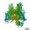
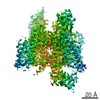
- Structure visualization
| Structure viewer | Molecule: MolmilJmol/JSmol |
|---|
- Downloads & links
-Download
| PDBx/mmCIF format | 6tk9.cif.gz | 313.1 KB | Display | PDBx/mmCIF format |
|---|---|---|---|---|
| PDB format | pdb6tk9.ent.gz | 256.1 KB | Display | PDB format |
| PDBx/mmJSON format | 6tk9.json.gz | Tree view | PDBx/mmJSON format | |
| Others |  Other downloads Other downloads |
-Validation report
| Arichive directory | https://data.pdbj.org/pub/pdb/validation_reports/tk/6tk9ftp://data.pdbj.org/pub/pdb/validation_reports/tk/6tk9 | HTTPS FTP |
|---|
-Related structure data
| Related structure data |  4uc0S S: Starting model for refinement |
|---|---|
| Similar structure data |

-Links
 PDBj
PDBj- Assembly
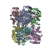
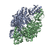
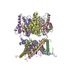
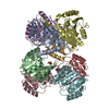
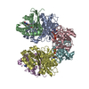
| Deposited unit | 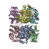
| ||||||||
|---|---|---|---|---|---|---|---|---|---|
| 1 |
| ||||||||
| Unit cell |
|
-Components
| #1: Protein | / Inosine-guanosine phosphorylase Mass: 29932.400 Da / Num. of mol.: 6 Source method: isolated from a genetically manipulated source Source: (gene. exp.) Thermus thermophilus (bacteria) / Gene: TthHC11_05990 / Production host: Thermus thermophilus (bacteria)References: UniProt: A0A510IGW9, purine-nucleoside phosphorylase#2: Chemical | ChemComp-PO4 / Phosphate  Mass: 94.971 Da / Num. of mol.: 6 / Source method: obtained synthetically / Formula: PO4 Mass: 94.971 Da / Num. of mol.: 6 / Source method: obtained synthetically / Formula: PO4#3: Water | ChemComp-HOH / | Water Mass: 18.015 Da / Num. of mol.: 57 / Source method: isolated from a natural source / Formula: H2O Mass: 18.015 Da / Num. of mol.: 57 / Source method: isolated from a natural source / Formula: H2OHas ligand of interest | N | |
|---|
-Experimental details
-Experiment
| Experiment | Method: X-RAY DIFFRACTION / Number of used crystals: 1 |
|---|
- Sample preparation
| Crystal | Density Matthews: 3.26 Å3/Da / Density % sol: 62.26 % |
|---|---|
| Crystal grow | Temperature: 297 K / Method: counter-diffusion / Details: SA |
-Data collection
| Diffraction | Mean temperature: 100 K / Serial crystal experiment: N |
|---|---|
| Diffraction source | Source: SYNCHROTRON / Site: ESRF  / Beamline: ID23-1 / Wavelength: 0.979 / Wavelength: 0.979 Å / Beamline: ID23-1 / Wavelength: 0.979 / Wavelength: 0.979 Å |
| Detector | Type: DECTRIS EIGER R 1M / Detector: PIXEL / Date: Oct 12, 2019 |
| Radiation | Protocol: SINGLE WAVELENGTH / Monochromatic (M) / Laue (L): M / Scattering type: x-ray |
| Radiation wavelength | Wavelength: 0.979 Å / Relative weight: 1 |
| Reflection | Resolution: 2.5→20 Å / Num. obs: 80416 / % possible obs: 98.1 % / Redundancy: 4.35 % / CC1/2: 0.998 / Rmerge(I) obs: 0.085 / Net I/σ(I): 13.32 |
| Reflection shell | Resolution: 2.5→3 Å / Rmerge(I) obs: 0.435 / Num. unique obs: 33938 / CC1/2: 0.922 / Rrim(I) all: 0.491 |
- Processing
| Software |
| ||||||||||||||||||||||||||||||||||||||||||||||||||||||||||||
|---|---|---|---|---|---|---|---|---|---|---|---|---|---|---|---|---|---|---|---|---|---|---|---|---|---|---|---|---|---|---|---|---|---|---|---|---|---|---|---|---|---|---|---|---|---|---|---|---|---|---|---|---|---|---|---|---|---|---|---|---|---|
| Refinement | Method to determine structure: MOLECULAR REPLACEMENT Starting model: 4UC0 Resolution: 2.5→19.98 Å / Cor.coef. Fo:Fc: 0.94 / Cor.coef. Fo:Fc free: 0.911 / SU B: 10.587 / SU ML: 0.225 / Cross valid method: THROUGHOUT / σ(F): 0 / ESU R: 0.395 / ESU R Free: 0.278 Details: HYDROGENS HAVE BEEN ADDED IN THE RIDING POSITIONS U VALUES : REFINED INDIVIDUALLY
| ||||||||||||||||||||||||||||||||||||||||||||||||||||||||||||
| Solvent computation | Ion probe radii: 0.8 Å / Shrinkage radii: 0.8 Å / VDW probe radii: 1.2 Å | ||||||||||||||||||||||||||||||||||||||||||||||||||||||||||||
| Displacement parameters | Biso max: 147.19 Å2 / Biso mean: 47.895 Å2 / Biso min: 21.22 Å2
| ||||||||||||||||||||||||||||||||||||||||||||||||||||||||||||
| Refinement step | Cycle: final / Resolution: 2.5→19.98 Å
| ||||||||||||||||||||||||||||||||||||||||||||||||||||||||||||
| Refine LS restraints |
| ||||||||||||||||||||||||||||||||||||||||||||||||||||||||||||
| LS refinement shell | Resolution: 2.5→2.564 Å / Rfactor Rfree error: 0 / Total num. of bins used: 20
|
