 Movie
Movie Controller

[English] 日本語
 Yorodumi
- PDB-4m1e: Crystal structure of purine nucleoside phosphorylase I from Planc... -
+ Open data

- Basic information
| Entry | Database: PDB / ID: 4m1e | ||||||
|---|---|---|---|---|---|---|---|
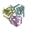
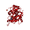
| Title | Crystal structure of purine nucleoside phosphorylase I from Planctomyces limnophilus DSM 3776, NYSGRC Target 029364. | ||||||
 Components Components | Purine nucleoside phosphorylase | ||||||
 Keywords Keywords | TRANSFERASE / STRUCTURAL GENOMICS / PROTEIN STRUCTURE INITIATIVE / NYSGRC / purine nucleoside phosphorylase / PSI-Biology / New York Structural Genomics Research Consortium | ||||||
| Function / homology |  Function and homology information Function and homology informationnucleoside metabolic process / purine-nucleoside phosphorylase / purine-nucleoside phosphorylase activitySimilarity search - Function | ||||||
| Biological species |  Planctomyces limnophilus (bacteria) Planctomyces limnophilus (bacteria) | ||||||
| Method | X-RAY DIFFRACTION / SYNCHROTRON / MOLECULAR REPLACEMENT / molecular replacement / Resolution: 1.9 Å | ||||||
 Authors Authors | Malashkevich, V.N. / Bonanno, J.B. / Bhosle, R. / Toro, R. / Hillerich, B. / Gizzi, A. / Garforth, S. / Kar, A. / Chan, M.K. / Lafluer, J. ...Malashkevich, V.N. / Bonanno, J.B. / Bhosle, R. / Toro, R. / Hillerich, B. / Gizzi, A. / Garforth, S. / Kar, A. / Chan, M.K. / Lafluer, J. / Patel, H. / Matikainen, B. / Chamala, S. / Lim, S. / Celikgil, A. / Villegas, G. / Evans, B. / Love, J. / Fiser, A. / Khafizov, K. / Seidel, R. / Almo, S.C. / New York Structural Genomics Research Consortium (NYSGRC) | ||||||
 Citation Citation | Journal: To be Published Title: Crystal structure of purine nucleoside phosphorylase I from Planctomyces limnophilus DSM 3776, NYSGRC Target 029364. Authors: Malashkevich, V.N. / Bonanno, J.B. / Bhosle, R. / Toro, R. / Hillerich, B. / Gizzi, A. / Garforth, S. / Kar, A. / Chan, M.K. / Lafluer, J. / Patel, H. / Matikainen, B. / Chamala, S. / Lim, S. ...Authors: Malashkevich, V.N. / Bonanno, J.B. / Bhosle, R. / Toro, R. / Hillerich, B. / Gizzi, A. / Garforth, S. / Kar, A. / Chan, M.K. / Lafluer, J. / Patel, H. / Matikainen, B. / Chamala, S. / Lim, S. / Celikgil, A. / Villegas, G. / Evans, B. / Love, J. / Fiser, A. / Khafizov, K. / Seidel, R. / Almo, S.C. | ||||||
| History |
|
- Structure visualization
| Structure viewer | Molecule: MolmilJmol/JSmol |
|---|
- Downloads & links
-Download
| PDBx/mmCIF format | 4m1e.cif.gz | 636.5 KB | Display | PDBx/mmCIF format |
|---|---|---|---|---|
| PDB format | pdb4m1e.ent.gz | 529.2 KB | Display | PDB format |
| PDBx/mmJSON format | 4m1e.json.gz | Tree view | PDBx/mmJSON format | |
| Others |  Other downloads Other downloads |
-Validation report
| Arichive directory | https://data.pdbj.org/pub/pdb/validation_reports/m1/4m1eftp://data.pdbj.org/pub/pdb/validation_reports/m1/4m1e | HTTPS FTP |
|---|
-Related structure data
| Related structure data |  3la8S S: Starting model for refinement |
|---|---|
| Similar structure data | |
| Other databases |


-Links
 PDBj
PDBj


- Assembly
| Deposited unit | 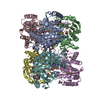
| ||||||||
|---|---|---|---|---|---|---|---|---|---|
| 1 | 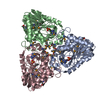
| ||||||||
| 2 | 
| ||||||||
| 3 |
| ||||||||
| Unit cell |
| ||||||||
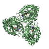
| Details | trimeric |
-Components
| #1: Protein | / Inosine-guanosine phosphorylase Mass: 32992.242 Da / Num. of mol.: 6 Source method: isolated from a genetically manipulated source Source: (gene. exp.) Planctomyces limnophilus (bacteria) / Strain: DSM 3776 / Gene: Plim_2216 / Plasmid: BC-PSGX3(BC) / Production host: Escherichia coli (E. coli) / Strain (production host): BL21(DE3)CODON+RILReferences: UniProt: D5SMY7, purine-nucleoside phosphorylase#2: Chemical | ChemComp-SO4 / Sulfate  Mass: 96.063 Da / Num. of mol.: 6 / Source method: obtained synthetically / Formula: SO4 Mass: 96.063 Da / Num. of mol.: 6 / Source method: obtained synthetically / Formula: SO4#3: Chemical | ChemComp-ADE / Adenine  Mass: 135.127 Da / Num. of mol.: 6 / Source method: obtained synthetically / Formula: C5H5N5 Mass: 135.127 Da / Num. of mol.: 6 / Source method: obtained synthetically / Formula: C5H5N5#4: Chemical | ChemComp-6PC / Picolinic acid  Mass: 123.109 Da / Num. of mol.: 6 / Source method: obtained synthetically / Formula: C6H5NO2 Mass: 123.109 Da / Num. of mol.: 6 / Source method: obtained synthetically / Formula: C6H5NO2#5: Water | ChemComp-HOH / | Water Mass: 18.015 Da / Num. of mol.: 662 / Source method: isolated from a natural source / Formula: H2O Mass: 18.015 Da / Num. of mol.: 662 / Source method: isolated from a natural source / Formula: H2ONonpolymer details | LIGAND PYRIDINE-2-CARBOXYLIC ACID IS EXOGENOUS AND WAS NOT ADDED TO THE CRYSTALLIZATION BUFFER. IT ...LIGAND PYRIDINE-2-CARBOXYLIC | |
|---|
-Experimental details
-Experiment
| Experiment | Method: X-RAY DIFFRACTION / Number of used crystals: 1 |
|---|
- Sample preparation
| Crystal | Density Matthews: 2.5 Å3/Da / Density % sol: 50.85 % |
|---|---|
| Crystal grow | Temperature: 298 K / Method: vapor diffusion, sitting drop / pH: 6.5 Details: 0.1 M MES:NaOH, pH 6.5, 1.6 Magnesium sulfate, VAPOR DIFFUSION, SITTING DROP, temperature 298K |
-Data collection
| Diffraction | Mean temperature: 100 K | |||||||||||||||||||||||||||||||||||||||||||||||||||||||||||||||||||||||||||||||||||||||||||||||||||||||||||||||||||||||||||||||||||||||||||||||||||
|---|---|---|---|---|---|---|---|---|---|---|---|---|---|---|---|---|---|---|---|---|---|---|---|---|---|---|---|---|---|---|---|---|---|---|---|---|---|---|---|---|---|---|---|---|---|---|---|---|---|---|---|---|---|---|---|---|---|---|---|---|---|---|---|---|---|---|---|---|---|---|---|---|---|---|---|---|---|---|---|---|---|---|---|---|---|---|---|---|---|---|---|---|---|---|---|---|---|---|---|---|---|---|---|---|---|---|---|---|---|---|---|---|---|---|---|---|---|---|---|---|---|---|---|---|---|---|---|---|---|---|---|---|---|---|---|---|---|---|---|---|---|---|---|---|---|---|---|---|
| Diffraction source | Source: SYNCHROTRON / Site: APS  / Beamline: 31-ID / Wavelength: 0.9791 Å / Beamline: 31-ID / Wavelength: 0.9791 Å | |||||||||||||||||||||||||||||||||||||||||||||||||||||||||||||||||||||||||||||||||||||||||||||||||||||||||||||||||||||||||||||||||||||||||||||||||||
| Detector | Type: RAYONIX MX225HE / Detector: CCD / Date: Jul 18, 2013 | |||||||||||||||||||||||||||||||||||||||||||||||||||||||||||||||||||||||||||||||||||||||||||||||||||||||||||||||||||||||||||||||||||||||||||||||||||
| Radiation | Protocol: SINGLE WAVELENGTH / Scattering type: x-ray | |||||||||||||||||||||||||||||||||||||||||||||||||||||||||||||||||||||||||||||||||||||||||||||||||||||||||||||||||||||||||||||||||||||||||||||||||||
| Radiation wavelength | Wavelength: 0.9791 Å / Relative weight: 1 | |||||||||||||||||||||||||||||||||||||||||||||||||||||||||||||||||||||||||||||||||||||||||||||||||||||||||||||||||||||||||||||||||||||||||||||||||||
| Reflection | Resolution: 1.9→50 Å / Num. obs: 148934 / % possible obs: 97 % / Redundancy: 2.1 % / Rmerge(I) obs: 0.113 / Χ2: 1.149 / Net I/σ(I): 9.7 | |||||||||||||||||||||||||||||||||||||||||||||||||||||||||||||||||||||||||||||||||||||||||||||||||||||||||||||||||||||||||||||||||||||||||||||||||||
| Reflection shell |
|
-Phasing
| Phasing | Method: molecular replacement | |||||||||
|---|---|---|---|---|---|---|---|---|---|---|
| Phasing MR | Model details: Phaser MODE: MR_AUTO
|
- Processing
| Software |
| |||||||||||||||||||||||||||||||||||||||||||||||||||||||||||||||||||||||||||||||||||||||||||||||||||||||||||||||||||||||||||||||||||||||||||||||||||||||||||||||||||||||||||||||
|---|---|---|---|---|---|---|---|---|---|---|---|---|---|---|---|---|---|---|---|---|---|---|---|---|---|---|---|---|---|---|---|---|---|---|---|---|---|---|---|---|---|---|---|---|---|---|---|---|---|---|---|---|---|---|---|---|---|---|---|---|---|---|---|---|---|---|---|---|---|---|---|---|---|---|---|---|---|---|---|---|---|---|---|---|---|---|---|---|---|---|---|---|---|---|---|---|---|---|---|---|---|---|---|---|---|---|---|---|---|---|---|---|---|---|---|---|---|---|---|---|---|---|---|---|---|---|---|---|---|---|---|---|---|---|---|---|---|---|---|---|---|---|---|---|---|---|---|---|---|---|---|---|---|---|---|---|---|---|---|---|---|---|---|---|---|---|---|---|---|---|---|---|---|---|---|---|
| Refinement | Method to determine structure: MOLECULAR REPLACEMENT Starting model: 3LA8 Resolution: 1.9→20 Å / Cor.coef. Fo:Fc: 0.939 / Cor.coef. Fo:Fc free: 0.912 / WRfactor Rfree: 0.2836 / WRfactor Rwork: 0.2337 / Occupancy max: 1 / Occupancy min: 0.5 / FOM work R set: 0.8176 / SU B: 8.152 / SU ML: 0.12 / SU R Cruickshank DPI: 0.163 / SU Rfree: 0.155 / Cross valid method: THROUGHOUT / σ(F): 0 / ESU R: 0.163 / ESU R Free: 0.155 / Stereochemistry target values: MAXIMUM LIKELIHOOD Details: U VALUES : WITH TLS ADDED HYDROGENS HAVE BEEN USED IF PRESENT IN THE INPUT
| |||||||||||||||||||||||||||||||||||||||||||||||||||||||||||||||||||||||||||||||||||||||||||||||||||||||||||||||||||||||||||||||||||||||||||||||||||||||||||||||||||||||||||||||
| Solvent computation | Ion probe radii: 0.8 Å / Shrinkage radii: 0.8 Å / VDW probe radii: 1.2 Å / Solvent model: MASK | |||||||||||||||||||||||||||||||||||||||||||||||||||||||||||||||||||||||||||||||||||||||||||||||||||||||||||||||||||||||||||||||||||||||||||||||||||||||||||||||||||||||||||||||
| Displacement parameters | Biso max: 145.21 Å2 / Biso mean: 34.4156 Å2 / Biso min: 13 Å2
| |||||||||||||||||||||||||||||||||||||||||||||||||||||||||||||||||||||||||||||||||||||||||||||||||||||||||||||||||||||||||||||||||||||||||||||||||||||||||||||||||||||||||||||||
| Refinement step | Cycle: LAST / Resolution: 1.9→20 Å
| |||||||||||||||||||||||||||||||||||||||||||||||||||||||||||||||||||||||||||||||||||||||||||||||||||||||||||||||||||||||||||||||||||||||||||||||||||||||||||||||||||||||||||||||
| Refine LS restraints |
| |||||||||||||||||||||||||||||||||||||||||||||||||||||||||||||||||||||||||||||||||||||||||||||||||||||||||||||||||||||||||||||||||||||||||||||||||||||||||||||||||||||||||||||||
| LS refinement shell | Resolution: 1.9→1.949 Å / Total num. of bins used: 20
| |||||||||||||||||||||||||||||||||||||||||||||||||||||||||||||||||||||||||||||||||||||||||||||||||||||||||||||||||||||||||||||||||||||||||||||||||||||||||||||||||||||||||||||||
| Refinement TLS params. | Method: refined / Refine-ID: X-RAY DIFFRACTION
| |||||||||||||||||||||||||||||||||||||||||||||||||||||||||||||||||||||||||||||||||||||||||||||||||||||||||||||||||||||||||||||||||||||||||||||||||||||||||||||||||||||||||||||||
| Refinement TLS group |
|
