 Movie
Movie Controller

[English] 日本語
 Yorodumi
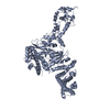
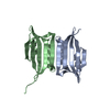
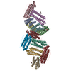
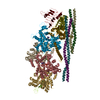
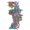
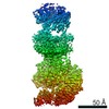
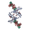
- PDB-6thh: Crystal structure of type I-D CRISPR-Cas nuclease Cas10d in compl... -
+ Open data

- Basic information
| Entry | Database: PDB / ID: 6thh | |||||||||
|---|---|---|---|---|---|---|---|---|---|---|
| Title | Crystal structure of type I-D CRISPR-Cas nuclease Cas10d in complex with the SIRV3 AcrID1 (gp02) anti-CRISPR protein | |||||||||
 Components Components |
| |||||||||
 Keywords Keywords |  IMMUNE SYSTEM / CRISPR / Cas / type I-D / Cas10 / Cas10d / complex / anti-CRISPR IMMUNE SYSTEM / CRISPR / Cas / type I-D / Cas10 / Cas10d / complex / anti-CRISPR | |||||||||
| Function / homology | Sulfolobus islandicus filamentous virus, Orf14 / Protein of unknown function (DUF1374) / PHOSPHATE ION / MafI family immunity protein / CRISPR-associated protein, CscA Function and homology information Function and homology information | |||||||||
| Biological species |   Sulfolobus islandicus rudivirus 3 Sulfolobus islandicus rudivirus 3 Sulfolobus islandicus LAL14/1 (acidophilic) Sulfolobus islandicus LAL14/1 (acidophilic) | |||||||||
| Method | X-RAY DIFFRACTION / SYNCHROTRON / SAD / Resolution: 3.48 Å | |||||||||
 Authors Authors | Manav, M.C. / Brodersen, D.E. | |||||||||
| Funding support |  Denmark, 2items Denmark, 2items
| |||||||||
 Citation Citation | Journal: Nat Commun / Year: 2020 Title: Structural basis for inhibition of an archaeal CRISPR-Cas type I-D large subunit by an anti-CRISPR protein. Authors: Manav, M.C. / Van, L.B. / Lin, J. / Fuglsang, A. / Peng, X. / Brodersen, D.E. | |||||||||
| History |
|
- Structure visualization
| Structure viewer | Molecule: MolmilJmol/JSmol |
|---|
- Downloads & links
-Download
| PDBx/mmCIF format | 6thh.cif.gz | 213.4 KB | Display | PDBx/mmCIF format |
|---|---|---|---|---|
| PDB format | pdb6thh.ent.gz | 176.2 KB | Display | PDB format |
| PDBx/mmJSON format | 6thh.json.gz | Tree view | PDBx/mmJSON format | |
| Others |  Other downloads Other downloads |
-Validation report
| Arichive directory | https://data.pdbj.org/pub/pdb/validation_reports/th/6thhftp://data.pdbj.org/pub/pdb/validation_reports/th/6thh | HTTPS FTP |
|---|
-Related structure data


-Links
 PDBj
PDBj
- Assembly
| Deposited unit | 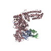
| ||||||||
|---|---|---|---|---|---|---|---|---|---|
| 1 | 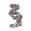
| ||||||||
| Unit cell |
|
-Components
| #1: Protein | Mass: 13086.693 Da / Num. of mol.: 2 Source method: isolated from a genetically manipulated source Source: (gene. exp.) Sulfolobus islandicus rudivirus 3 / Production host:  Escherichia coli (E. coli) / References: UniProt: A0A1B3SN05 Escherichia coli (E. coli) / References: UniProt: A0A1B3SN05#2: Protein | | Mass: 98332.102 Da / Num. of mol.: 1 Source method: isolated from a genetically manipulated source Source: (gene. exp.) Sulfolobus islandicus LAL14/1 (acidophilic)Gene: SiL_0609 / Production host: Escherichia coli (E. coli) / References: UniProt: M9U4Y8#3: Chemical | ChemComp-PO4 / | Phosphate  Mass: 94.971 Da / Num. of mol.: 1 / Source method: obtained synthetically / Formula: PO4 / Feature type: SUBJECT OF INVESTIGATION Mass: 94.971 Da / Num. of mol.: 1 / Source method: obtained synthetically / Formula: PO4 / Feature type: SUBJECT OF INVESTIGATION#4: Chemical | ChemComp-ZN / |   Mass: 65.409 Da / Num. of mol.: 1 / Source method: obtained synthetically / Formula: Zn / Feature type: SUBJECT OF INVESTIGATION Mass: 65.409 Da / Num. of mol.: 1 / Source method: obtained synthetically / Formula: Zn / Feature type: SUBJECT OF INVESTIGATIONHas ligand of interest | Y | |
|---|
-Experimental details
-Experiment
| Experiment | Method: X-RAY DIFFRACTION / Number of used crystals: 1 |
|---|
- Sample preparation
| Crystal | Density Matthews: 3.37 Å3/Da / Density % sol: 67 % |
|---|---|
| Crystal grow | Temperature: 292 K / Method: vapor diffusion, sitting drop / pH: 7.5 Details: 0.1 M Hepes 7.5, 2% (w/v) PEG 8000, 5% ethylene glycol |
-Data collection
| Diffraction | Mean temperature: 100 K / Serial crystal experiment: N |
|---|---|
| Diffraction source | Source: SYNCHROTRON / Site: PETRA III, EMBL c/o DESY  / Beamline: P14 (MX2) / Wavelength: 0.9793 Å / Beamline: P14 (MX2) / Wavelength: 0.9793 Å |
| Detector | Type: DECTRIS EIGER X 16M / Detector: PIXEL / Date: Sep 20, 2019 |
| Radiation | Protocol: SINGLE WAVELENGTH / Monochromatic (M) / Laue (L): M / Scattering type: x-ray |
| Radiation wavelength | Wavelength: 0.9793 Å / Relative weight: 1 |
| Reflection | Resolution: 3.48→67.542 Å / Num. obs: 21713 / % possible obs: 99.9 % / Redundancy: 2 % / Biso Wilson estimate: 137.1 Å2 / CC1/2: 0.99 / Rmerge(I) obs: 0.06 / Net I/σ(I): 11.9 |
| Reflection shell | Resolution: 3.48→3.6 Å / Redundancy: 2 % / Rmerge(I) obs: 0.68 / Mean I/σ(I) obs: 1.2 / Num. unique obs: 2139 / CC1/2: 0.6 / % possible all: 99.8 |
-Phasing
| Phasing | Method: SAD |
|---|
- Processing
| Software |
| ||||||||||||||||||||||||
|---|---|---|---|---|---|---|---|---|---|---|---|---|---|---|---|---|---|---|---|---|---|---|---|---|---|
| Refinement | Method to determine structure: SAD / Resolution: 3.48→67.542 Å / SU ML: 0.46 / Cross valid method: THROUGHOUT / σ(F): 1.34 / Phase error: 26.77 / Stereochemistry target values: ML
| ||||||||||||||||||||||||
| Solvent computation | Shrinkage radii: 0.9 Å / VDW probe radii: 1.11 Å / Solvent model: FLAT BULK SOLVENT MODEL | ||||||||||||||||||||||||
| Displacement parameters | Biso max: 282.81 Å2 / Biso mean: 137.0532 Å2 / Biso min: 61.23 Å2 | ||||||||||||||||||||||||
| Refinement step | Cycle: final / Resolution: 3.48→67.542 Å
| ||||||||||||||||||||||||
| LS refinement shell |
|
