 Movie
Movie Controller

[English] 日本語
 Yorodumi
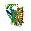
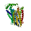
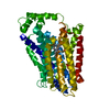
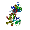
- PDB-6tha: Crystal structure of human sugar transporter GLUT1 (SLC2A1) in th... -
+ Open data

- Basic information
| Entry | Database: PDB / ID: 6tha | ||||||||||||
|---|---|---|---|---|---|---|---|---|---|---|---|---|---|
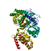
| Title | Crystal structure of human sugar transporter GLUT1 (SLC2A1) in the inward conformation | ||||||||||||
 Components Components | Solute carrier family 2, facilitated glucose transporter member 1 | ||||||||||||
 Keywords Keywords | MEMBRANE PROTEIN / ALPHA-HELICAL PROTEIN / SUGAR TRANSPORT | ||||||||||||
| Function / homology |  Function and homology informationglucose transporter complex / Defective SLC2A1 causes GLUT1 deficiency syndrome 1 (GLUT1DS1) / long-chain fatty acid import across plasma membrane / response to Thyroglobulin triiodothyronine / dehydroascorbic acid transmembrane transporter activity / dehydroascorbic acid transport / Lactose synthesis / glucose transmembrane transporter activity / D-glucose transmembrane transporter activity / Cellular hexose transport ...glucose transporter complex / Defective SLC2A1 causes GLUT1 deficiency syndrome 1 (GLUT1DS1) / long-chain fatty acid import across plasma membrane / response to Thyroglobulin triiodothyronine / dehydroascorbic acid transmembrane transporter activity / dehydroascorbic acid transport / Lactose synthesis / glucose transmembrane transporter activity / D-glucose transmembrane transporter activity / Cellular hexose transport / Vitamin C (ascorbate) metabolism / glucose import across plasma membrane / L-ascorbic acid metabolic process / glucose transmembrane transport / cellular hyperosmotic response / photoreceptor cell maintenance / female germ cell nucleus / female pronucleus / long-chain fatty acid transmembrane transporter activity / cortical actin cytoskeleton / glucose import / xenobiotic transmembrane transporter activity / transport across blood-brain barrier / intercalated disc / cellular response to glucose starvation / photoreceptor inner segment / central nervous system development / female pregnancy / Regulation of insulin secretion / caveola / sarcolemma / response to insulin / cerebral cortex development / Z disc / kinase binding / cellular response to mechanical stimulus / : / melanosome / presynapse / midbody / basolateral plasma membrane / protein-containing complex assembly / blood microparticle / response to hypoxia / apical plasma membrane / Golgi membrane / extracellular exosome / membrane / identical protein binding / plasma membrane / cytosol Function and homology informationglucose transporter complex / Defective SLC2A1 causes GLUT1 deficiency syndrome 1 (GLUT1DS1) / long-chain fatty acid import across plasma membrane / response to Thyroglobulin triiodothyronine / dehydroascorbic acid transmembrane transporter activity / dehydroascorbic acid transport / Lactose synthesis / glucose transmembrane transporter activity / D-glucose transmembrane transporter activity / Cellular hexose transport ...glucose transporter complex / Defective SLC2A1 causes GLUT1 deficiency syndrome 1 (GLUT1DS1) / long-chain fatty acid import across plasma membrane / response to Thyroglobulin triiodothyronine / dehydroascorbic acid transmembrane transporter activity / dehydroascorbic acid transport / Lactose synthesis / glucose transmembrane transporter activity / D-glucose transmembrane transporter activity / Cellular hexose transport / Vitamin C (ascorbate) metabolism / glucose import across plasma membrane / L-ascorbic acid metabolic process / glucose transmembrane transport / cellular hyperosmotic response / photoreceptor cell maintenance / female germ cell nucleus / female pronucleus / long-chain fatty acid transmembrane transporter activity / cortical actin cytoskeleton / glucose import / xenobiotic transmembrane transporter activity / transport across blood-brain barrier / intercalated disc / cellular response to glucose starvation / photoreceptor inner segment / central nervous system development / female pregnancy / Regulation of insulin secretion / caveola / sarcolemma / response to insulin / cerebral cortex development / Z disc / kinase binding / cellular response to mechanical stimulus / : / melanosome / presynapse / midbody / basolateral plasma membrane / protein-containing complex assembly / blood microparticle / response to hypoxia / apical plasma membrane / Golgi membrane / extracellular exosome / membrane / identical protein binding / plasma membrane / cytosolSimilarity search - Function | ||||||||||||
| Biological species |  Homo sapiens (human) Homo sapiens (human) | ||||||||||||
| Method | X-RAY DIFFRACTION / SYNCHROTRON / MOLECULAR REPLACEMENT / molecular replacement / Resolution: 2.4 Å | ||||||||||||
 Authors Authors | Pedersen, B.P. / Paulsen, P.A. / Custodio, T.F. | ||||||||||||
| Funding support |  Denmark, 3items Denmark, 3items
| ||||||||||||
 Citation Citation | Journal: Life Sci Alliance / Year: 2021 Title: Structural comparison of GLUT1 to GLUT3 reveal transport regulation mechanism in sugar porter family. Authors: Custodio, T.F. / Paulsen, P.A. / Frain, K.M. / Pedersen, B.P. | ||||||||||||
| History |
|
- Structure visualization
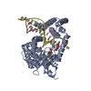
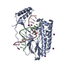
| Structure viewer | Molecule: MolmilJmol/JSmol |
|---|
- Downloads & links
-Download
| PDBx/mmCIF format | 6tha.cif.gz | 193.7 KB | Display | PDBx/mmCIF format |
|---|---|---|---|---|
| PDB format | pdb6tha.ent.gz | 154.4 KB | Display | PDB format |
| PDBx/mmJSON format | 6tha.json.gz | Tree view | PDBx/mmJSON format | |
| Others |  Other downloads Other downloads |
-Validation report
| Arichive directory | https://data.pdbj.org/pub/pdb/validation_reports/th/6thaftp://data.pdbj.org/pub/pdb/validation_reports/th/6tha | HTTPS FTP |
|---|
-Related structure data
| Related structure data | 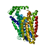 4pypS S: Starting model for refinement |
|---|---|
| Similar structure data |


-Links
 PDBj
PDBj


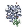
- Assembly
| Deposited unit | 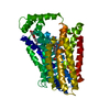
| ||||||||
|---|---|---|---|---|---|---|---|---|---|
| 1 |
| ||||||||
| Unit cell |
|
-Components
| #1: Protein | / Glucose transporter type 1 / erythrocyte/brain / GLUT-1 / HepG2 glucose transporter Mass: 54591.992 Da / Num. of mol.: 1 Source method: isolated from a genetically manipulated source Source: (gene. exp.) Homo sapiens (human) / Gene: SLC2A1, GLUT1 / Production host:  Saccharomyces cerevisiae (brewer's yeast) / References: UniProt: P11166 Saccharomyces cerevisiae (brewer's yeast) / References: UniProt: P11166 | ||||||||
|---|---|---|---|---|---|---|---|---|---|
| #2: Sugar | 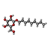  Type: D-saccharide / Mass: 306.395 Da / Num. of mol.: 2 Type: D-saccharide / Mass: 306.395 Da / Num. of mol.: 2Source method: isolated from a genetically manipulated source Formula: C15H30O6 / Feature type: SUBJECT OF INVESTIGATION / Comment: detergent*YM #3: Chemical | ChemComp-P33 / | Polyethylene glycol  Mass: 326.383 Da / Num. of mol.: 1 / Source method: obtained synthetically / Formula: C14H30O8 / Comment: precipitant*YM Mass: 326.383 Da / Num. of mol.: 1 / Source method: obtained synthetically / Formula: C14H30O8 / Comment: precipitant*YM#4: Chemical | ChemComp-CL / | Chloride  Mass: 35.453 Da / Num. of mol.: 1 / Source method: obtained synthetically / Formula: Cl / Feature type: SUBJECT OF INVESTIGATION Mass: 35.453 Da / Num. of mol.: 1 / Source method: obtained synthetically / Formula: Cl / Feature type: SUBJECT OF INVESTIGATION#5: Water | ChemComp-HOH / | Water Mass: 18.015 Da / Num. of mol.: 13 / Source method: isolated from a natural source / Formula: H2O Mass: 18.015 Da / Num. of mol.: 13 / Source method: isolated from a natural source / Formula: H2OHas ligand of interest | Y | |
-Experimental details
-Experiment
| Experiment | Method: X-RAY DIFFRACTION / Number of used crystals: 1 |
|---|
- Sample preparation
| Crystal | Density Matthews: 3.83 Å3/Da / Density % sol: 67.87 % |
|---|---|
| Crystal grow | Temperature: 290.15 K / Method: vapor diffusion, hanging drop Details: 42-46 % polyethylene glycol 400, 100-200 mM MgCl2 and 100 mM Mops pH 7.4 |
-Data collection
| Diffraction | Mean temperature: 100 K / Serial crystal experiment: N | ||||||||||||||||||||||||||||||||||||||||||||||||||||||||||||||||||||||||||||||||||||||||||||||||||||||||||||||||||||||||||||||||||||||||||||||||||||||||
|---|---|---|---|---|---|---|---|---|---|---|---|---|---|---|---|---|---|---|---|---|---|---|---|---|---|---|---|---|---|---|---|---|---|---|---|---|---|---|---|---|---|---|---|---|---|---|---|---|---|---|---|---|---|---|---|---|---|---|---|---|---|---|---|---|---|---|---|---|---|---|---|---|---|---|---|---|---|---|---|---|---|---|---|---|---|---|---|---|---|---|---|---|---|---|---|---|---|---|---|---|---|---|---|---|---|---|---|---|---|---|---|---|---|---|---|---|---|---|---|---|---|---|---|---|---|---|---|---|---|---|---|---|---|---|---|---|---|---|---|---|---|---|---|---|---|---|---|---|---|---|---|---|---|
| Diffraction source | Source: SYNCHROTRON / Site: Diamond  / Beamline: I24 / Wavelength: 0.9686 Å / Beamline: I24 / Wavelength: 0.9686 Å | ||||||||||||||||||||||||||||||||||||||||||||||||||||||||||||||||||||||||||||||||||||||||||||||||||||||||||||||||||||||||||||||||||||||||||||||||||||||||
| Detector | Type: DECTRIS PILATUS3 R CdTe 300K / Detector: PIXEL / Date: Feb 5, 2017 | ||||||||||||||||||||||||||||||||||||||||||||||||||||||||||||||||||||||||||||||||||||||||||||||||||||||||||||||||||||||||||||||||||||||||||||||||||||||||
| Radiation | Protocol: SINGLE WAVELENGTH / Monochromatic (M) / Laue (L): M / Scattering type: x-ray | ||||||||||||||||||||||||||||||||||||||||||||||||||||||||||||||||||||||||||||||||||||||||||||||||||||||||||||||||||||||||||||||||||||||||||||||||||||||||
| Radiation wavelength | Wavelength: 0.9686 Å / Relative weight: 1 | ||||||||||||||||||||||||||||||||||||||||||||||||||||||||||||||||||||||||||||||||||||||||||||||||||||||||||||||||||||||||||||||||||||||||||||||||||||||||
| Reflection | Resolution: 2.4→41.656 Å / Num. obs: 32166 / % possible obs: 99.9 % / Redundancy: 7.145 % / Biso Wilson estimate: 78.65 Å2 / CC1/2: 0.998 / Rmerge(I) obs: 0.113 / Rrim(I) all: 0.123 / Χ2: 1.163 / Net I/σ(I): 7.2 / Num. measured all: 229827 / Scaling rejects: 993 | ||||||||||||||||||||||||||||||||||||||||||||||||||||||||||||||||||||||||||||||||||||||||||||||||||||||||||||||||||||||||||||||||||||||||||||||||||||||||
| Reflection shell | Diffraction-ID: 1
|
-Phasing
| Phasing | Method: molecular replacement |
|---|
- Processing
| Software |
| ||||||||||||||||||||||||||||||||||||||||||||||||||||||||||||||||||||||||||||||||||||||||||||||||||||
|---|---|---|---|---|---|---|---|---|---|---|---|---|---|---|---|---|---|---|---|---|---|---|---|---|---|---|---|---|---|---|---|---|---|---|---|---|---|---|---|---|---|---|---|---|---|---|---|---|---|---|---|---|---|---|---|---|---|---|---|---|---|---|---|---|---|---|---|---|---|---|---|---|---|---|---|---|---|---|---|---|---|---|---|---|---|---|---|---|---|---|---|---|---|---|---|---|---|---|---|---|---|
| Refinement | Method to determine structure: MOLECULAR REPLACEMENT Starting model: 4PYP Resolution: 2.4→41.656 Å / SU ML: 0.47 / Cross valid method: THROUGHOUT / σ(F): 1.34 / Phase error: 34.13 / Stereochemistry target values: ML
| ||||||||||||||||||||||||||||||||||||||||||||||||||||||||||||||||||||||||||||||||||||||||||||||||||||
| Solvent computation | Shrinkage radii: 0.9 Å / VDW probe radii: 1.11 Å / Solvent model: FLAT BULK SOLVENT MODEL | ||||||||||||||||||||||||||||||||||||||||||||||||||||||||||||||||||||||||||||||||||||||||||||||||||||
| Displacement parameters | Biso max: 206.76 Å2 / Biso mean: 105.9745 Å2 / Biso min: 57.33 Å2 | ||||||||||||||||||||||||||||||||||||||||||||||||||||||||||||||||||||||||||||||||||||||||||||||||||||
| Refinement step | Cycle: final / Resolution: 2.4→41.656 Å
| ||||||||||||||||||||||||||||||||||||||||||||||||||||||||||||||||||||||||||||||||||||||||||||||||||||
| Refine LS restraints |
| ||||||||||||||||||||||||||||||||||||||||||||||||||||||||||||||||||||||||||||||||||||||||||||||||||||
| LS refinement shell | Refine-ID: X-RAY DIFFRACTION / Rfactor Rfree error: 0
| ||||||||||||||||||||||||||||||||||||||||||||||||||||||||||||||||||||||||||||||||||||||||||||||||||||
| Refinement TLS params. | Method: refined / Refine-ID: X-RAY DIFFRACTION
| ||||||||||||||||||||||||||||||||||||||||||||||||||||||||||||||||||||||||||||||||||||||||||||||||||||
| Refinement TLS group |
|
