 Movie
Movie Controller

[English] 日本語
 Yorodumi
- PDB-6tez: Crystal Structure of full-length Human Lysyl Hydroxylase LH3 - Va... -
+ Open data

- Basic information
| Entry | Database: PDB / ID: 6tez | |||||||||||||||
|---|---|---|---|---|---|---|---|---|---|---|---|---|---|---|---|---|
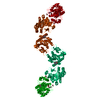
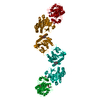
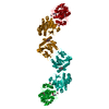
| Title | Crystal Structure of full-length Human Lysyl Hydroxylase LH3 - Val80Lys mutant - Cocrystal with Fe2+, Mn2+, UDP-Glucuronic Acid | |||||||||||||||
 Components Components | Multifunctional procollagen lysine hydroxylase and glycosyltransferase LH3 | |||||||||||||||
 Keywords Keywords |  TRANSFERASE / Collagen / lysyl hydroxylase / hydroxylysine / galactosyltransferase / glucosyltransferase TRANSFERASE / Collagen / lysyl hydroxylase / hydroxylysine / galactosyltransferase / glucosyltransferase | |||||||||||||||
| Function / homology |  Function and homology informationprocollagen galactosyltransferase / procollagen glucosyltransferase / peptidyl-lysine hydroxylation / procollagen glucosyltransferase activity / hydroxylysine biosynthetic process / procollagen galactosyltransferase activity / procollagen-lysine 5-dioxygenase / procollagen-lysine 5-dioxygenase activity / basement membrane assembly / epidermis morphogenesis ...procollagen galactosyltransferase / procollagen glucosyltransferase / peptidyl-lysine hydroxylation / procollagen glucosyltransferase activity / hydroxylysine biosynthetic process / procollagen galactosyltransferase activity / procollagen-lysine 5-dioxygenase / procollagen-lysine 5-dioxygenase activity / basement membrane assembly / epidermis morphogenesis / Collagen biosynthesis and modifying enzymes / collagen metabolic process / endothelial cell morphogenesis / protein O-linked glycosylation / L-ascorbic acid binding / collagen fibril organization / neural tube development / lung morphogenesis / small molecule binding / rough endoplasmic reticulum / trans-Golgi network / protein localization / vasodilation / collagen-containing extracellular matrix / in utero embryonic development / iron ion binding / endoplasmic reticulum lumen / endoplasmic reticulum membrane / Golgi apparatus / endoplasmic reticulum / extracellular space / extracellular exosome / metal ion binding Function and homology informationprocollagen galactosyltransferase / procollagen glucosyltransferase / peptidyl-lysine hydroxylation / procollagen glucosyltransferase activity / hydroxylysine biosynthetic process / procollagen galactosyltransferase activity / procollagen-lysine 5-dioxygenase / procollagen-lysine 5-dioxygenase activity / basement membrane assembly / epidermis morphogenesis ...procollagen galactosyltransferase / procollagen glucosyltransferase / peptidyl-lysine hydroxylation / procollagen glucosyltransferase activity / hydroxylysine biosynthetic process / procollagen galactosyltransferase activity / procollagen-lysine 5-dioxygenase / procollagen-lysine 5-dioxygenase activity / basement membrane assembly / epidermis morphogenesis / Collagen biosynthesis and modifying enzymes / collagen metabolic process / endothelial cell morphogenesis / protein O-linked glycosylation / L-ascorbic acid binding / collagen fibril organization / neural tube development / lung morphogenesis / small molecule binding / rough endoplasmic reticulum / trans-Golgi network / protein localization / vasodilation / collagen-containing extracellular matrix / in utero embryonic development / iron ion binding / endoplasmic reticulum lumen / endoplasmic reticulum membrane / Golgi apparatus / endoplasmic reticulum / extracellular space / extracellular exosome / metal ion bindingSimilarity search - Function | |||||||||||||||
| Biological species |  Homo sapiens (human) Homo sapiens (human) | |||||||||||||||
| Method | X-RAY DIFFRACTION / SYNCHROTRON / MOLECULAR REPLACEMENT / Resolution: 2.7 Å | |||||||||||||||
 Authors Authors | Chiapparino, A. / De Giorgi, F. / Scietti, L. / Faravelli, S. / Roscioli, T. / Forneris, F. | |||||||||||||||
| Funding support |  Italy, 4items Italy, 4items
| |||||||||||||||
 Citation Citation | Journal: Int J Mol Sci / Year: 2023 Title: Identification of Regulatory Molecular 'Hot Spots' for LH/PLOD Collagen Glycosyltransferase Activity Authors: Mattoteia, D. / Chiapparino, A. / Fumagalli, M. / De Marco, M. / De Giorgi, F. / Negro, L. / Pinnola, A. / Faravelli, S. / Roscioli, T. / Scietti, L. / Forneris, F. | |||||||||||||||
| History |
|
- Structure visualization
| Structure viewer | Molecule: MolmilJmol/JSmol |
|---|
- Downloads & links
-Download
| PDBx/mmCIF format | 6tez.cif.gz | 362.5 KB | Display | PDBx/mmCIF format |
|---|---|---|---|---|
| PDB format | pdb6tez.ent.gz | 244.9 KB | Display | PDB format |
| PDBx/mmJSON format | 6tez.json.gz | Tree view | PDBx/mmJSON format | |
| Others |  Other downloads Other downloads |
-Validation report
| Arichive directory | https://data.pdbj.org/pub/pdb/validation_reports/te/6tezftp://data.pdbj.org/pub/pdb/validation_reports/te/6tez | HTTPS FTP |
|---|
-Related structure data
| Related structure data |  6te3C  6tecC  6tesC  6teuC  6texC  6fxkS S: Starting model for refinement C: citing same article ( |
|---|---|
| Similar structure data |


-Links
 PDBj
PDBj


- Assembly
| Deposited unit | 
| ||||||||||||
|---|---|---|---|---|---|---|---|---|---|---|---|---|---|
| 1 | 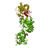
| ||||||||||||
| Unit cell |
| ||||||||||||
| Components on special symmetry positions |
|
-Components
-Protein , 1 types, 1 molecules A
| #1: Protein | Mass: 82814.461 Da / Num. of mol.: 1 Source method: isolated from a genetically manipulated source Details: Val80Lys Mutation - First Ser residue and last three Ala residues were introduced by molecular cloning Source: (gene. exp.) Homo sapiens (human) / Gene: PLOD3 / Cell line (production host): HEK293F / Production host: Homo sapiens (human)References: UniProt: O60568, procollagen-lysine 5-dioxygenase, procollagen galactosyltransferase, procollagen glucosyltransferase |
|---|
-Sugars , 2 types, 2 molecules 
| #2: Polysaccharide | 2-acetamido-2-deoxy-beta-D-glucopyranose-(1-4)-2-acetamido-2-deoxy-beta-D-glucopyranose / Mass: 424.401 Da / Num. of mol.: 1 Source method: isolated from a genetically manipulated source |
|---|---|
| #8: Sugar | ChemComp-NAG / N-Acetylglucosamine Type: D-saccharide, beta linking / Mass: 221.208 Da / Num. of mol.: 1 Type: D-saccharide, beta linking / Mass: 221.208 Da / Num. of mol.: 1Source method: isolated from a genetically manipulated source Formula: C8H15NO6 |
-Non-polymers , 6 types, 80 molecules 


| #3: Chemical | ChemComp-UGA / Uridine diphosphate glucuronic acid Mass: 580.285 Da / Num. of mol.: 1 / Source method: obtained synthetically / Formula: C15H22N2O18P2 / Feature type: SUBJECT OF INVESTIGATION Mass: 580.285 Da / Num. of mol.: 1 / Source method: obtained synthetically / Formula: C15H22N2O18P2 / Feature type: SUBJECT OF INVESTIGATION | ||||||||
|---|---|---|---|---|---|---|---|---|---|
| #4: Chemical | Glycerol Mass: 92.094 Da / Num. of mol.: 2 / Source method: obtained synthetically / Formula: C3H8O3 Mass: 92.094 Da / Num. of mol.: 2 / Source method: obtained synthetically / Formula: C3H8O3#5: Chemical | ChemComp-AKG / | Α-Ketoglutaric acid Mass: 146.098 Da / Num. of mol.: 1 / Source method: obtained synthetically / Formula: C5H6O5 / Feature type: SUBJECT OF INVESTIGATION Mass: 146.098 Da / Num. of mol.: 1 / Source method: obtained synthetically / Formula: C5H6O5 / Feature type: SUBJECT OF INVESTIGATION#6: Chemical |  Mass: 55.845 Da / Num. of mol.: 2 / Source method: obtained synthetically / Formula: Fe / Feature type: SUBJECT OF INVESTIGATION Mass: 55.845 Da / Num. of mol.: 2 / Source method: obtained synthetically / Formula: Fe / Feature type: SUBJECT OF INVESTIGATION#7: Chemical | ChemComp-MN / |  Mass: 54.938 Da / Num. of mol.: 1 / Source method: obtained synthetically / Formula: Mn / Feature type: SUBJECT OF INVESTIGATION Mass: 54.938 Da / Num. of mol.: 1 / Source method: obtained synthetically / Formula: Mn / Feature type: SUBJECT OF INVESTIGATION#9: Water | ChemComp-HOH / | WaterMass: 18.015 Da / Num. of mol.: 73 / Source method: isolated from a natural source / Formula: H2O |
-Details
| Has ligand of interest | Y |
|---|
-Experimental details
-Experiment
| Experiment | Method: X-RAY DIFFRACTION / Number of used crystals: 1 |
|---|
- Sample preparation
| Crystal | Density Matthews: 3.31 Å3/Da / Density % sol: 62.87 % |
|---|---|
| Crystal grow | Temperature: 277 K / Method: vapor diffusion, hanging drop / pH: 7.8 Details: 600 mM sodium formate, 12% PGA-LM, 100 mM HEPES/NaOH, 500 uM FeCl2, 500 uM MnCl2, 1 mM UDP-Glucuronic Acid Temp details: cold room |
-Data collection
| Diffraction | Mean temperature: 100 K / Serial crystal experiment: N |
|---|---|
| Diffraction source | Source: SYNCHROTRON / Site: SLS  / Beamline: X06SA / Wavelength: 1 Å / Beamline: X06SA / Wavelength: 1 Å |
| Detector | Type: DECTRIS EIGER X 16M / Detector: PIXEL / Date: Nov 28, 2018 |
| Radiation | Protocol: SINGLE WAVELENGTH / Monochromatic (M) / Laue (L): M / Scattering type: x-ray |
| Radiation wavelength | Wavelength: 1 Å / Relative weight: 1 |
| Reflection | Resolution: 2.7→49 Å / Num. obs: 30477 / % possible obs: 99.5 % / Redundancy: 6.5 % / Biso Wilson estimate: 41.43 Å2 / CC1/2: 0.996 / Rmerge(I) obs: 0.138 / Rpim(I) all: 0.088 / Net I/σ(I): 9.4 |
| Reflection shell | Resolution: 2.7→2.83 Å / Rmerge(I) obs: 1.312 / Mean I/σ(I) obs: 1.3 / Num. unique obs: 3911 / CC1/2: 0.58 / Rpim(I) all: 0.902 |
- Processing
| Software |
| ||||||||||||||||||||||||||||||||||||||||||||||||||||||||||||||||||||||||||||||||||||||||||||||||||||
|---|---|---|---|---|---|---|---|---|---|---|---|---|---|---|---|---|---|---|---|---|---|---|---|---|---|---|---|---|---|---|---|---|---|---|---|---|---|---|---|---|---|---|---|---|---|---|---|---|---|---|---|---|---|---|---|---|---|---|---|---|---|---|---|---|---|---|---|---|---|---|---|---|---|---|---|---|---|---|---|---|---|---|---|---|---|---|---|---|---|---|---|---|---|---|---|---|---|---|---|---|---|
| Refinement | Method to determine structure: MOLECULAR REPLACEMENT Starting model: 6FXK Resolution: 2.7→49 Å / SU ML: 0.3007 / Cross valid method: FREE R-VALUE / σ(F): 1.35 / Phase error: 23.2911
| ||||||||||||||||||||||||||||||||||||||||||||||||||||||||||||||||||||||||||||||||||||||||||||||||||||
| Solvent computation | Shrinkage radii: 0.9 Å / VDW probe radii: 1.11 Å | ||||||||||||||||||||||||||||||||||||||||||||||||||||||||||||||||||||||||||||||||||||||||||||||||||||
| Displacement parameters | Biso mean: 43.73 Å2 | ||||||||||||||||||||||||||||||||||||||||||||||||||||||||||||||||||||||||||||||||||||||||||||||||||||
| Refinement step | Cycle: LAST / Resolution: 2.7→49 Å
| ||||||||||||||||||||||||||||||||||||||||||||||||||||||||||||||||||||||||||||||||||||||||||||||||||||
| Refine LS restraints |
| ||||||||||||||||||||||||||||||||||||||||||||||||||||||||||||||||||||||||||||||||||||||||||||||||||||
| LS refinement shell |
| ||||||||||||||||||||||||||||||||||||||||||||||||||||||||||||||||||||||||||||||||||||||||||||||||||||
| Refinement TLS params. | Method: refined / Refine-ID: X-RAY DIFFRACTION
| ||||||||||||||||||||||||||||||||||||||||||||||||||||||||||||||||||||||||||||||||||||||||||||||||||||
| Refinement TLS group |
|
