[English] 日本語
Yorodumi
- PDB-6fxm: Crystal Structure of full-length Human Lysyl Hydroxylase LH3 - Co... -

+
Open data


ID or keywords:

Loading...

-
Basic information

Entry
Database: PDB / ID: 6fxm
TitleCrystal Structure of full-length Human Lysyl Hydroxylase LH3 - Cocrystal with Mn2+
ComponentsProcollagen-lysine,2-oxoglutarate 5-dioxygenase 3
KeywordsTRANSFERASE / Lysyl Hydroxylase / Galacosyltransferase / Glucosyltransferase / Collagen
Function / homology
Function and homology information


procollagen galactosyltransferase / procollagen glucosyltransferase / peptidyl-lysine hydroxylation / procollagen glucosyltransferase activity / hydroxylysine biosynthetic process / procollagen galactosyltransferase activity / procollagen-lysine 5-dioxygenase / procollagen-lysine 5-dioxygenase activity / basement membrane assembly / epidermis morphogenesis ...procollagen galactosyltransferase / procollagen glucosyltransferase / peptidyl-lysine hydroxylation / procollagen glucosyltransferase activity / hydroxylysine biosynthetic process / procollagen galactosyltransferase activity / procollagen-lysine 5-dioxygenase / procollagen-lysine 5-dioxygenase activity / basement membrane assembly / epidermis morphogenesis / Collagen biosynthesis and modifying enzymes / collagen metabolic process / endothelial cell morphogenesis / protein O-linked glycosylation / L-ascorbic acid binding / collagen fibril organization / neural tube development / lung morphogenesis / small molecule binding / rough endoplasmic reticulum / trans-Golgi network / protein localization / vasodilation / collagen-containing extracellular matrix / in utero embryonic development / iron ion binding / endoplasmic reticulum lumen / endoplasmic reticulum membrane / Golgi apparatus / endoplasmic reticulum / extracellular space / extracellular exosome / metal ion binding
Similarity search - Function
Procollagen-lysine 5-dioxygenase, conserved site / Lysyl hydroxylase signature. / Prolyl 4-hydroxylase, alpha subunit / Prolyl 4-hydroxylase alpha subunit homologues. / Isopenicillin N synthase-like, Fe(2+) 2OG dioxygenase domain / 2OG-Fe(II) oxygenase superfamily / Oxoglutarate/iron-dependent dioxygenase / Fe(2+) 2-oxoglutarate dioxygenase domain profile. / Nucleotide-diphospho-sugar transferases
Similarity search - Domain/homology
2-OXOGLUTARIC ACID / : / : / Multifunctional procollagen lysine hydroxylase and glycosyltransferase LH3
Similarity search - Component
Biological speciesHomo sapiens (human)
MethodX-RAY DIFFRACTION / SYNCHROTRON / SAD / Resolution: 2.1 Å
AuthorsScietti, L. / Chiapparino, A. / De Giorgi, F. / Fumagalli, M. / Khoriauli, L. / Nergadze, S. / Basu, S. / Olieric, V. / Banushi, B. / Giulotto, E. ...Scietti, L. / Chiapparino, A. / De Giorgi, F. / Fumagalli, M. / Khoriauli, L. / Nergadze, S. / Basu, S. / Olieric, V. / Banushi, B. / Giulotto, E. / Gissen, P. / Forneris, F.
Funding support United States, Italy, 5items
OrganizationGrant numberCountry
The Giovanni Armenise-Harvard FoundationCareer Development Award United States
Cariplo Foundation2015-0768 - COME TRUE Italy
AIRC20075 Italy
European Commission MSCA-IF745934 - COTETHERS Italy
European Union - BIOSTRUCT7551, 10205 Italy
Citation
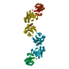
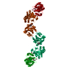
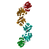
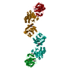
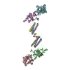
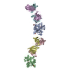
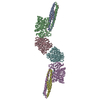
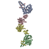
Journal: Nat Commun / Year: 2018
Title: Molecular architecture of the multifunctional collagen lysyl hydroxylase and glycosyltransferase LH3.
Authors: Luigi Scietti / Antonella Chiapparino / Francesca De Giorgi / Marco Fumagalli / Lela Khoriauli / Solomon Nergadze / Shibom Basu / Vincent Olieric / Lucia Cucca / Blerida Banushi / Antonella ...Authors: Luigi Scietti / Antonella Chiapparino / Francesca De Giorgi / Marco Fumagalli / Lela Khoriauli / Solomon Nergadze / Shibom Basu / Vincent Olieric / Lucia Cucca / Blerida Banushi / Antonella Profumo / Elena Giulotto / Paul Gissen / Federico Forneris /
Abstract: Lysyl hydroxylases catalyze hydroxylation of collagen lysines, and sustain essential roles in extracellular matrix (ECM) maturation and remodeling. Malfunctions in these enzymes cause severe ...Lysyl hydroxylases catalyze hydroxylation of collagen lysines, and sustain essential roles in extracellular matrix (ECM) maturation and remodeling. Malfunctions in these enzymes cause severe connective tissue disorders. Human lysyl hydroxylase 3 (LH3/PLOD3) bears multiple enzymatic activities, as it catalyzes collagen lysine hydroxylation and also their subsequent glycosylation. Our understanding of LH3 functions is currently hampered by lack of molecular structure information. Here, we present high resolution crystal structures of full-length human LH3 in complex with cofactors and donor substrates. The elongated homodimeric LH3 architecture shows two distinct catalytic sites at the N- and C-terminal boundaries of each monomer, separated by an accessory domain. The glycosyltransferase domain displays distinguishing features compared to other known glycosyltransferases. Known disease-related mutations map in close proximity to the catalytic sites. Collectively, our results provide a structural framework characterizing the multiple functions of LH3, and the molecular mechanisms of collagen-related diseases involving human lysyl hydroxylases.
History
DepositionMar 9, 2018Deposition site: PDBE / Processing site: PDBE
Revision 1.0Aug 22, 2018Provider: repository / Type: Initial release
Revision 2.0Jul 29, 2020Group: Atomic model / Data collection ...Atomic model / Data collection / Derived calculations / Structure summary
Category: atom_site / chem_comp ...atom_site / chem_comp / entity / pdbx_branch_scheme / pdbx_chem_comp_identifier / pdbx_entity_branch / pdbx_entity_branch_descriptor / pdbx_entity_branch_link / pdbx_entity_branch_list / pdbx_entity_nonpoly / pdbx_nonpoly_scheme / pdbx_struct_assembly_gen / pdbx_struct_conn_angle / pdbx_struct_special_symmetry / struct_asym / struct_conn / struct_site / struct_site_gen
Item: _atom_site.B_iso_or_equiv / _atom_site.Cartn_x ..._atom_site.B_iso_or_equiv / _atom_site.Cartn_x / _atom_site.Cartn_y / _atom_site.Cartn_z / _atom_site.auth_asym_id / _atom_site.auth_atom_id / _atom_site.auth_comp_id / _atom_site.auth_seq_id / _atom_site.label_asym_id / _atom_site.label_atom_id / _atom_site.label_comp_id / _atom_site.label_entity_id / _atom_site.type_symbol / _chem_comp.name / _chem_comp.type / _pdbx_entity_nonpoly.entity_id / _pdbx_entity_nonpoly.name / _pdbx_struct_assembly_gen.asym_id_list / _pdbx_struct_conn_angle.ptnr1_label_asym_id / _pdbx_struct_conn_angle.ptnr2_label_asym_id / _pdbx_struct_conn_angle.ptnr3_label_asym_id / _pdbx_struct_special_symmetry.label_asym_id / _struct_conn.conn_type_id / _struct_conn.id / _struct_conn.pdbx_dist_value / _struct_conn.pdbx_leaving_atom_flag / _struct_conn.pdbx_role / _struct_conn.ptnr1_auth_asym_id / _struct_conn.ptnr1_auth_comp_id / _struct_conn.ptnr1_auth_seq_id / _struct_conn.ptnr1_label_asym_id / _struct_conn.ptnr1_label_atom_id / _struct_conn.ptnr1_label_comp_id / _struct_conn.ptnr1_label_seq_id / _struct_conn.ptnr2_auth_asym_id / _struct_conn.ptnr2_auth_comp_id / _struct_conn.ptnr2_auth_seq_id / _struct_conn.ptnr2_label_asym_id / _struct_conn.ptnr2_label_atom_id / _struct_conn.ptnr2_label_comp_id
Description: Carbohydrate remediation / Provider: repository / Type: Remediation

-
Structure visualization

Structure viewerMolecule:
MolmilJmol/JSmol

Downloads & links

-
Assembly

Deposited unit
A: Procollagen-lysine,2-oxoglutarate 5-dioxygenase 3
hetero molecules


Theoretical massNumber of molelcules
Total (without water)84,11011
Polymers82,7841
Non-polymers1,32610
Water4,738263
1
A: Procollagen-lysine,2-oxoglutarate 5-dioxygenase 3
hetero molecules

A: Procollagen-lysine,2-oxoglutarate 5-dioxygenase 3
hetero molecules


  • defined by author
  • Evidence: SAXS, SEC-SAXS data highlight presence of dimers in solution, gel filtration, gel filtration data highlight presence of dimers in solution, homology, homologous viral structures (PDB ID ...Evidence: SAXS, SEC-SAXS data highlight presence of dimers in solution, gel filtration, gel filtration data highlight presence of dimers in solution, homology, homologous viral structures (PDB ID 6AX6 and 6AX7) show identical dimer interfaces
  • 168 kDa, 2 polymers
  • Search similar-shape structures of this assembly by Omokage search (details)
Theoretical massNumber of molelcules
Total (without water)168,22022
Polymers165,5692
Non-polymers2,65220
Water362
TypeNameSymmetry operationNumber
identity operation1_555x,y,z1
crystal symmetry operation4_557x,-y,-z+21
Unit cell
Length a, b, c (Å)97.270, 99.949, 225.138
Angle α, β, γ (deg.)90.00, 90.00, 90.00
Int Tables number20
Space group name H-MC2221
Components on special symmetry positions
IDModelComponents
11A-985-

HOH

21A-1094-

HOH

-
Components

-
Protein , 1 types, 1 molecules A

#1: Protein Procollagen-lysine,2-oxoglutarate 5-dioxygenase 3 / Lysyl hydroxylase 3 / LH3


Mass: 82784.406 Da / Num. of mol.: 1
Source method: isolated from a genetically manipulated source
Source: (gene. exp.) Homo sapiens (human) / Gene: PLOD3 / Plasmid: pPuro-DHFR / Cell (production host): HeLa / Production host: Homo sapiens (human)
References: UniProt: O60568, procollagen-lysine 5-dioxygenase

-
Sugars , 2 types, 2 molecules

#2: Polysaccharide 2-acetamido-2-deoxy-beta-D-glucopyranose-(1-4)-2-acetamido-2-deoxy-beta-D-glucopyranose


Type: oligosaccharide / Mass: 424.401 Da / Num. of mol.: 1
Source method: isolated from a genetically manipulated source
DescriptorTypeProgram
DGlcpNAcb1-4DGlcpNAcb1-Glycam Condensed SequenceGMML 1.0
WURCS=2.0/1,2,1/[a2122h-1b_1-5_2*NCC/3=O]/1-1/a4-b1WURCSPDB2Glycan 1.1.0
[]{[(4+1)][b-D-GlcpNAc]{[(4+1)][b-D-GlcpNAc]{}}}LINUCSPDB-CARE
#7: Sugar ChemComp-NAG / 2-acetamido-2-deoxy-beta-D-glucopyranose / N-Acetylglucosamine


Type: D-saccharide, beta linking / Mass: 221.208 Da / Num. of mol.: 1
Source method: isolated from a genetically manipulated source
Formula: C8H15NO6
IdentifierTypeProgram
DGlcpNAcbCONDENSED IUPAC CARBOHYDRATE SYMBOLGMML 1.0
N-acetyl-b-D-glucopyranosamineCOMMON NAMEGMML 1.0
b-D-GlcpNAcIUPAC CARBOHYDRATE SYMBOLPDB-CARE 1.0
GlcNAcSNFG CARBOHYDRATE SYMBOLGMML 1.0

-
Non-polymers , 5 types, 271 molecules

#3: Chemical
ChemComp-GOL / GLYCEROL / GLYCERIN / PROPANE-1,2,3-TRIOL / Glycerol


Mass: 92.094 Da / Num. of mol.: 4 / Source method: obtained synthetically / Formula: C3H8O3
#4: Chemical ChemComp-AKG / 2-OXOGLUTARIC ACID / Α-Ketoglutaric acid


Mass: 146.098 Da / Num. of mol.: 1 / Source method: obtained synthetically / Formula: C5H6O5 / Feature type: SUBJECT OF INVESTIGATION
#5: Chemical ChemComp-FE2 / FE (II) ION


Mass: 55.845 Da / Num. of mol.: 1 / Source method: obtained synthetically / Formula: Fe / Feature type: SUBJECT OF INVESTIGATION
#6: Chemical ChemComp-MN / MANGANESE (II) ION


Mass: 54.938 Da / Num. of mol.: 2 / Source method: obtained synthetically / Formula: Mn / Feature type: SUBJECT OF INVESTIGATION
#8: Water ChemComp-HOH / water / Water


Mass: 18.015 Da / Num. of mol.: 263 / Source method: isolated from a natural source / Formula: H2O

-
Experimental details

-
Experiment

ExperimentMethod: X-RAY DIFFRACTION / Number of used crystals: 1

-
Sample preparation

CrystalDensity Matthews: 3.1 Å3/Da / Density % sol: 60 %
Crystal growTemperature: 277 K / Method: vapor diffusion, hanging drop / pH: 7.8
Details: 600 mM sodium formate, 12% PGA-LM, 100 mM HEPES/NaOH, 0.5 mM MnCl2
Temp details: cold room

-
Data collection

DiffractionMean temperature: 100 K
Diffraction sourceSource: SYNCHROTRON / Site: ESRF / Beamline: MASSIF-3 / Wavelength: 0.9677 Å
DetectorType: DECTRIS EIGER X 4M / Detector: PIXEL / Date: Apr 11, 2017
RadiationProtocol: SINGLE WAVELENGTH / Monochromatic (M) / Laue (L): M / Scattering type: x-ray
Radiation wavelengthWavelength: 0.9677 Å / Relative weight: 1
ReflectionResolution: 2.1→48.79 Å / Num. obs: 64193 / % possible obs: 99.8 % / Redundancy: 7.8 % / CC1/2: 0.999 / Rmerge(I) obs: 0.095 / Rpim(I) all: 0.054 / Net I/σ(I): 12.2
Reflection shellResolution: 2.1→2.15 Å / Redundancy: 8 % / Rmerge(I) obs: 2.444 / Mean I/σ(I) obs: 0.9 / Num. unique obs: 4455 / Rpim(I) all: 1.389 / % possible all: 99.8

-
Processing

Software
NameVersionClassification
PHENIX(1.10.1_2155: ???)refinement
XDSdata reduction
Aimlessdata scaling
SHARPphasing
RefinementMethod to determine structure: SAD / Resolution: 2.1→37.37 Å / SU ML: 0.31 / Cross valid method: FREE R-VALUE / σ(F): 1.34 / Phase error: 39.86
RfactorNum. reflection% reflectionSelection details
Rfree0.2518 3132 4.9 %Random selection
Rwork0.2288 ---
obs0.2299 63945 99.52 %-
Solvent computationShrinkage radii: 0.9 Å / VDW probe radii: 1.11 Å
Refinement stepCycle: LAST / Resolution: 2.1→37.37 Å
ProteinNucleic acidLigandSolventTotal
Num. atoms5555 0 79 263 5897
Refine LS restraints
Refine-IDTypeDev idealNumber
X-RAY DIFFRACTIONf_bond_d0.0035811
X-RAY DIFFRACTIONf_angle_d0.6187894
X-RAY DIFFRACTIONf_dihedral_angle_d15.4753433
X-RAY DIFFRACTIONf_chiral_restr0.044830
X-RAY DIFFRACTIONf_plane_restr0.0041025
LS refinement shell
Resolution (Å)Rfactor RfreeNum. reflection RfreeRfactor RworkNum. reflection RworkRefine-ID% reflection obs (%)
2.1-2.13280.52811490.5412718X-RAY DIFFRACTION99
2.1328-2.16780.61621400.51042737X-RAY DIFFRACTION99
2.1678-2.20520.54461260.47532731X-RAY DIFFRACTION100
2.2052-2.24530.48921390.472769X-RAY DIFFRACTION100
2.2453-2.28840.45531330.43582706X-RAY DIFFRACTION99
2.2884-2.33520.44251350.40382765X-RAY DIFFRACTION100
2.3352-2.38590.38261370.36922735X-RAY DIFFRACTION100
2.3859-2.44140.37191420.34542735X-RAY DIFFRACTION99
2.4414-2.50250.36571440.34182756X-RAY DIFFRACTION99
2.5025-2.57010.38831310.33242752X-RAY DIFFRACTION99
2.5701-2.64570.31041650.28742690X-RAY DIFFRACTION99
2.6457-2.73110.34291310.28192763X-RAY DIFFRACTION100
2.7311-2.82870.31661620.26582741X-RAY DIFFRACTION100
2.8287-2.94190.26011510.23822753X-RAY DIFFRACTION100
2.9419-3.07570.25961440.22762766X-RAY DIFFRACTION100
3.0757-3.23780.23931290.21742783X-RAY DIFFRACTION100
3.2378-3.44050.23861270.20472772X-RAY DIFFRACTION99
3.4405-3.70590.21751410.19082798X-RAY DIFFRACTION100
3.7059-4.07850.19271550.16912756X-RAY DIFFRACTION99
4.0785-4.66770.16891490.14452806X-RAY DIFFRACTION100
4.6677-5.87710.19251580.16012835X-RAY DIFFRACTION100
5.8771-37.37580.16591440.17882946X-RAY DIFFRACTION100
Refinement TLS params.

Method: refined / Refine-ID: X-RAY DIFFRACTION

IDL112)L122)L132)L222)L232)L332)S11 (Å °)S12 (Å °)S13 (Å °)S21 (Å °)S22 (Å °)S23 (Å °)S31 (Å °)S32 (Å °)S33 (Å °)T112)T122)T132)T222)T232)T332)Origin x (Å)Origin y (Å)Origin z (Å)
10.01670.00820.00150.0130.01880.0192-0.22130.1583-0.0622-0.15570.0246-0.2277-0.35450.0866-0.00030.53830.0314-0.0260.34150.00170.315970.558730.7908146.8295
20.4590.3287-0.01570.8157-0.05120.2245-0.10290.1258-0.2814-0.3364-0.0153-0.4079-0.2822-0.4963-0.10730.11630.2032-0.0930.4624-0.08080.131957.469822.9206145.7471
30.00420.0367-0.02690.1041-0.04220.08540.06320.0737-0.09060.133-0.0105-0.1121-0.04830.00080.02470.17240.0315-0.0490.2722-0.03840.282971.489125.8466170.1193
40.29060.11690.02010.135-0.09380.15770.0652-0.00620.0310.2702-0.0372-0.0071-0.02870.01530.00030.2534-0.0243-0.0280.19870.01150.304772.050535.7841183.0134
50.1203-0.0142-0.00590.1239-0.04230.1338-0.03070.0170.01960.176-0.02070.0199-0.03880.0827-0.00050.629-0.0956-0.04870.36960.04250.263577.968918.3114219.2569
Refinement TLS group
IDRefine-IDRefine TLS-IDSelection details
1X-RAY DIFFRACTION1chain 'A' and (resid 33 through 100 )
2X-RAY DIFFRACTION2chain 'A' and (resid 101 through 242 )
3X-RAY DIFFRACTION3chain 'A' and (resid 243 through 310 )
4X-RAY DIFFRACTION4chain 'A' and (resid 311 through 515 )
5X-RAY DIFFRACTION5chain 'A' and (resid 516 through 738 )

+
About Yorodumi

-
News

-
Feb 9, 2022. New format data for meta-information of EMDB entries

New format data for meta-information of EMDB entries

  • Version 3 of the EMDB header file is now the official format.
  • The previous official version 1.9 will be removed from the archive.

Related info.:EMDB header

External links:wwPDB to switch to version 3 of the EMDB data model

-
Aug 12, 2020. Covid-19 info

Covid-19 info

URL: https://pdbj.org/emnavi/covid19.php

New page: Covid-19 featured information page in EM Navigator.

Related info.:Covid-19 info / Mar 5, 2020. Novel coronavirus structure data

+
Mar 5, 2020. Novel coronavirus structure data

Novel coronavirus structure data

Related info.:Yorodumi Speices / Aug 12, 2020. Covid-19 info

External links:COVID-19 featured content - PDBj / Molecule of the Month (242):Coronavirus Proteases

+
Jan 31, 2019. EMDB accession codes are about to change! (news from PDBe EMDB page)

EMDB accession codes are about to change! (news from PDBe EMDB page)

  • The allocation of 4 digits for EMDB accession codes will soon come to an end. Whilst these codes will remain in use, new EMDB accession codes will include an additional digit and will expand incrementally as the available range of codes is exhausted. The current 4-digit format prefixed with “EMD-” (i.e. EMD-XXXX) will advance to a 5-digit format (i.e. EMD-XXXXX), and so on. It is currently estimated that the 4-digit codes will be depleted around Spring 2019, at which point the 5-digit format will come into force.
  • The EM Navigator/Yorodumi systems omit the EMD- prefix.

Related info.:Q: What is EMD? / ID/Accession-code notation in Yorodumi/EM Navigator

External links:EMDB Accession Codes are Changing Soon! / Contact to PDBj

+
Jul 12, 2017. Major update of PDB

Major update of PDB

  • wwPDB released updated PDB data conforming to the new PDBx/mmCIF dictionary.
  • This is a major update changing the version number from 4 to 5, and with Remediation, in which all the entries are updated.
  • In this update, many items about electron microscopy experimental information are reorganized (e.g. em_software).
  • Now, EM Navigator and Yorodumi are based on the updated data.

External links:wwPDB Remediation / Enriched Model Files Conforming to OneDep Data Standards Now Available in the PDB FTP Archive

-
Yorodumi

Thousand views of thousand structures

  • Yorodumi is a browser for structure data from EMDB, PDB, SASBDB, etc.
  • This page is also the successor to EM Navigator detail page, and also detail information page/front-end page for Omokage search.
  • The word "yorodu" (or yorozu) is an old Japanese word meaning "ten thousand". "mi" (miru) is to see.

Related info.:EMDB / PDB / SASBDB / Comparison of 3 databanks / Yorodumi Search / Aug 31, 2016. New EM Navigator & Yorodumi / Yorodumi Papers / Jmol/JSmol / Function and homology information / Changes in new EM Navigator and Yorodumi

Read more