 Movie
Movie Controller

[English] 日本語
 Yorodumi
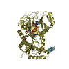
- PDB-6t8l: Crystal structure of Bacteroides thetaiotamicron EndoBT-3987 with... -
+ Open data

- Basic information
| Entry | Database: PDB / ID: 6t8l | |||||||||
|---|---|---|---|---|---|---|---|---|---|---|
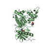
| Title | Crystal structure of Bacteroides thetaiotamicron EndoBT-3987 with Man9GlcNAc product in P212121 | |||||||||
 Components Components | Endo-beta-N-acetylglucosaminidase F1 | |||||||||
 Keywords Keywords |  HYDROLASE / endo-b-N-acetylglucosaminidase / EndoBT / glycoside hydrolase HYDROLASE / endo-b-N-acetylglucosaminidase / EndoBT / glycoside hydrolase | |||||||||
| Function / homology |  Function and homology information Function and homology information | |||||||||
| Biological species |  Bacteroides thetaiotaomicron VPI-5482 (bacteria) Bacteroides thetaiotaomicron VPI-5482 (bacteria) | |||||||||
| Method | X-RAY DIFFRACTION / SYNCHROTRON / MOLECULAR REPLACEMENT / Resolution: 1.7 Å | |||||||||
 Authors Authors | Trastoy, B. / Du, J.J. / Klontz, E.H. / Cifuente, J.O. / Sundberg, E.J. / Guerin, M.E. | |||||||||
 Citation Citation | Journal: Nat Commun / Year: 2020 Title: Structural basis of mammalian high-mannose N-glycan processing by human gut Bacteroides. Authors: Trastoy, B. / Du, J.J. / Klontz, E.H. / Li, C. / Cifuente, J.O. / Wang, L.X. / Sundberg, E.J. / Guerin, M.E. | |||||||||
| History |
|
- Structure visualization
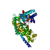
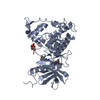
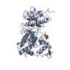
| Structure viewer | Molecule: MolmilJmol/JSmol |
|---|
- Downloads & links
-Download
| PDBx/mmCIF format | 6t8l.cif.gz | 201 KB | Display | PDBx/mmCIF format |
|---|---|---|---|---|
| PDB format | pdb6t8l.ent.gz | 156.1 KB | Display | PDB format |
| PDBx/mmJSON format | 6t8l.json.gz | Tree view | PDBx/mmJSON format | |
| Others |  Other downloads Other downloads |
-Validation report
| Arichive directory | https://data.pdbj.org/pub/pdb/validation_reports/t8/6t8lftp://data.pdbj.org/pub/pdb/validation_reports/t8/6t8l | HTTPS FTP |
|---|
-Related structure data
| Related structure data | 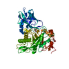 6t8iC 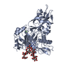 6t8kC 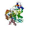 6tcvC 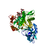 6tcwC 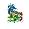 3pohS S: Starting model for refinement C: citing same article ( |
|---|---|
| Similar structure data |

-Links
 PDBj
PDBj


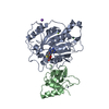
- Assembly
| Deposited unit | 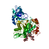
| ||||||||
|---|---|---|---|---|---|---|---|---|---|
| 1 |
| ||||||||
| Unit cell |
|
-Components
| #1: Protein | Mass: 49193.746 Da / Num. of mol.: 1 Source method: isolated from a genetically manipulated source Source: (gene. exp.) Bacteroides thetaiotaomicron VPI-5482 (bacteria)Gene: BT_3987 / Production host: Escherichia coli (E. coli) / References: UniProt: Q8A0N4 |
|---|---|
| #2: Polysaccharide | alpha-D-mannopyranose-(1-2)-alpha-D-mannopyranose-(1-2)-alpha-D-mannopyranose-(1-3)-[alpha-D- ...alpha-D-mannopyranose-(1-2)-alpha-D-mannopyranose-(1-2)-alpha-D-mannopyranose-(1-3)-[alpha-D-mannopyranose-(1-2)-alpha-D-mannopyranose-(1-3)-[alpha-D-mannopyranose-(1-2)-alpha-D-mannopyranose-(1-6)]alpha-D-mannopyranose-(1-6)]beta-D-mannopyranose-(1-4)-2-acetamido-2-deoxy-beta-D-glucopyranose / Mass: 1680.475 Da / Num. of mol.: 1 Source method: isolated from a genetically manipulated source |
| #3: Chemical | ChemComp-CA /   Mass: 40.078 Da / Num. of mol.: 1 / Source method: obtained synthetically / Formula: Ca Mass: 40.078 Da / Num. of mol.: 1 / Source method: obtained synthetically / Formula: Ca |
| #4: Water | ChemComp-HOH / Water Mass: 18.015 Da / Num. of mol.: 375 / Source method: isolated from a natural source / Formula: H2O Mass: 18.015 Da / Num. of mol.: 375 / Source method: isolated from a natural source / Formula: H2O |
| Has ligand of interest | Y |
-Experimental details
-Experiment
| Experiment | Method: X-RAY DIFFRACTION / Number of used crystals: 1 |
|---|
- Sample preparation
| Crystal | Density Matthews: 2.23 Å3/Da / Density % sol: 44.96 % |
|---|---|
| Crystal grow | Temperature: 291 K / Method: vapor diffusion, sitting drop Details: 0.02 M sodium/potassium phosphate, 100 mM Bis-Tris propane pH 8.5, and 20% (w/v) PEG 3350 |
-Data collection
| Diffraction | Mean temperature: 100 K / Serial crystal experiment: N |
|---|---|
| Diffraction source | Source: SYNCHROTRON / Site: Diamond  / Beamline: I03 / Wavelength: 0.97624 Å / Beamline: I03 / Wavelength: 0.97624 Å |
| Detector | Type: DECTRIS PILATUS 6M / Detector: PIXEL / Date: Dec 9, 2018 |
| Radiation | Protocol: SINGLE WAVELENGTH / Monochromatic (M) / Laue (L): M / Scattering type: x-ray |
| Radiation wavelength | Wavelength: 0.97624 Å / Relative weight: 1 |
| Reflection | Resolution: 1.7→29.178 Å / Num. obs: 49300 / % possible obs: 99.96 % / Redundancy: 6.6 % / CC1/2: 0.999 / Rmerge(I) obs: 0.08 / Net I/σ(I): 16.46 |
| Reflection shell | Resolution: 1.7→29.178 Å / Rmerge(I) obs: 0.482 / Num. unique obs: 4854 / CC1/2: 0.903 |
- Processing
| Software |
| |||||||||||||||||||||||||||||||||||||||||||||||||||||||||||||||||||||||||||||||||||||||||||||||
|---|---|---|---|---|---|---|---|---|---|---|---|---|---|---|---|---|---|---|---|---|---|---|---|---|---|---|---|---|---|---|---|---|---|---|---|---|---|---|---|---|---|---|---|---|---|---|---|---|---|---|---|---|---|---|---|---|---|---|---|---|---|---|---|---|---|---|---|---|---|---|---|---|---|---|---|---|---|---|---|---|---|---|---|---|---|---|---|---|---|---|---|---|---|---|---|---|
| Refinement | Method to determine structure: MOLECULAR REPLACEMENT Starting model: 3POH Resolution: 1.7→29.178 Å / SU ML: 0.14 / Cross valid method: THROUGHOUT / σ(F): 1.34 / Phase error: 17.49 / Stereochemistry target values: ML
| |||||||||||||||||||||||||||||||||||||||||||||||||||||||||||||||||||||||||||||||||||||||||||||||
| Solvent computation | Shrinkage radii: 0.9 Å / VDW probe radii: 1.11 Å / Solvent model: FLAT BULK SOLVENT MODEL | |||||||||||||||||||||||||||||||||||||||||||||||||||||||||||||||||||||||||||||||||||||||||||||||
| Displacement parameters | Biso max: 55.93 Å2 / Biso mean: 18.4092 Å2 / Biso min: 8.56 Å2 | |||||||||||||||||||||||||||||||||||||||||||||||||||||||||||||||||||||||||||||||||||||||||||||||
| Refinement step | Cycle: final / Resolution: 1.7→29.178 Å
| |||||||||||||||||||||||||||||||||||||||||||||||||||||||||||||||||||||||||||||||||||||||||||||||
| LS refinement shell | Refine-ID: X-RAY DIFFRACTION / Rfactor Rfree error: 0 / % reflection obs: 100 %
| |||||||||||||||||||||||||||||||||||||||||||||||||||||||||||||||||||||||||||||||||||||||||||||||
| Refinement TLS params. | Method: refined / Origin x: 4.3783 Å / Origin y: 19.7825 Å / Origin z: 26.8334 Å
| |||||||||||||||||||||||||||||||||||||||||||||||||||||||||||||||||||||||||||||||||||||||||||||||
| Refinement TLS group |
|
