 Movie
Movie Controller

[English] 日本語
 Yorodumi
- PDB-6t2t: Crystal structure of Drosophila melanogaster glutathione S-transf... -
+ Open data

- Basic information
| Entry | Database: PDB / ID: 6t2t | ||||||||||||
|---|---|---|---|---|---|---|---|---|---|---|---|---|---|
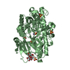
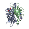
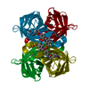
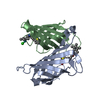
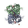
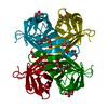
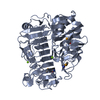
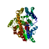
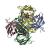
| Title | Crystal structure of Drosophila melanogaster glutathione S-transferase epsilon 14 in complex with glutathione and 2-methyl-2,4-pentanediol | ||||||||||||
 Components Components | Glutathione S-transferase E14 | ||||||||||||
 Keywords Keywords |  TRANSFERASE / glutathione transferase / steroid isomerase TRANSFERASE / glutathione transferase / steroid isomerase | ||||||||||||
| Function / homology |  Function and homology information Function and homology informationecdysteroid biosynthetic process / steroid delta-isomerase activity / glutathione transferase / glutathione transferase activity / glutathione metabolic process / cholesterol homeostasis / response to toxic substance / cytoplasmSimilarity search - Function | ||||||||||||
| Biological species |  Drosophila melanogaster (fruit fly) Drosophila melanogaster (fruit fly) | ||||||||||||
| Method | X-RAY DIFFRACTION / SYNCHROTRON / MOLECULAR REPLACEMENT / molecular replacement / Resolution: 1.3 Å | ||||||||||||
 Authors Authors | Skerlova, J. / Lindstrom, H. / Sjodin, B. / Gonis, E. / Neiers, F. / Mannervik, B. / Stenmark, P. | ||||||||||||
| Funding support |  Czech Republic, Czech Republic,  Sweden, 3items Sweden, 3items
| ||||||||||||
 Citation Citation | Journal: Febs Lett. / Year: 2020 Title: Structure and steroid isomerase activity of Drosophila glutathione transferase E14 essential for ecdysteroid biosynthesis. Authors: Skerlova, J. / Lindstrom, H. / Gonis, E. / Sjodin, B. / Neiers, F. / Stenmark, P. / Mannervik, B. | ||||||||||||
| History |
|
- Structure visualization
| Structure viewer | Molecule: MolmilJmol/JSmol |
|---|
- Downloads & links
-Download
| PDBx/mmCIF format | 6t2t.cif.gz | 70.8 KB | Display | PDBx/mmCIF format |
|---|---|---|---|---|
| PDB format | pdb6t2t.ent.gz | 50.8 KB | Display | PDB format |
| PDBx/mmJSON format | 6t2t.json.gz | Tree view | PDBx/mmJSON format | |
| Others |  Other downloads Other downloads |
-Validation report
| Arichive directory | https://data.pdbj.org/pub/pdb/validation_reports/t2/6t2tftp://data.pdbj.org/pub/pdb/validation_reports/t2/6t2t | HTTPS FTP |
|---|
-Related structure data
| Related structure data | 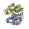 3vwxS S: Starting model for refinement |
|---|---|
| Similar structure data |

-Links
 PDBj
PDBj

- Assembly
| Deposited unit | 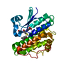
| ||||||||||||
|---|---|---|---|---|---|---|---|---|---|---|---|---|---|
| 1 | 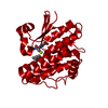
| ||||||||||||
| Unit cell |
| ||||||||||||
| Components on special symmetry positions |
|
-Components
| #1: Protein | Mass: 27479.492 Da / Num. of mol.: 1 Source method: isolated from a genetically manipulated source Source: (gene. exp.) Drosophila melanogaster (fruit fly) / Gene: GstE14, GSTD14-14, nobo, CG4688 / Production host:  Escherichia coli (E. coli) / References: UniProt: Q7JYX0, glutathione transferase Escherichia coli (E. coli) / References: UniProt: Q7JYX0, glutathione transferase |
|---|---|
| #2: Chemical | ChemComp-GSH / Glutathione  Mass: 307.323 Da / Num. of mol.: 1 / Source method: obtained synthetically / Formula: C10H17N3O6S / Feature type: SUBJECT OF INVESTIGATION Mass: 307.323 Da / Num. of mol.: 1 / Source method: obtained synthetically / Formula: C10H17N3O6S / Feature type: SUBJECT OF INVESTIGATION |
| #3: Chemical | ChemComp-MPD / (2-Methyl-2,4-pentanediol  Mass: 118.174 Da / Num. of mol.: 1 / Source method: obtained synthetically / Formula: C6H14O2 / Feature type: SUBJECT OF INVESTIGATION / Comment: precipitant*YM Mass: 118.174 Da / Num. of mol.: 1 / Source method: obtained synthetically / Formula: C6H14O2 / Feature type: SUBJECT OF INVESTIGATION / Comment: precipitant*YM |
| #4: Water | ChemComp-HOH / Water Mass: 18.015 Da / Num. of mol.: 237 / Source method: isolated from a natural source / Formula: H2O Mass: 18.015 Da / Num. of mol.: 237 / Source method: isolated from a natural source / Formula: H2O |
| Has ligand of interest | Y |
-Experimental details
-Experiment
| Experiment | Method: X-RAY DIFFRACTION / Number of used crystals: 1 |
|---|
- Sample preparation
| Crystal | Density Matthews: 2.66 Å3/Da / Density % sol: 53.73 % / Description: cubic |
|---|---|
| Crystal grow | Temperature: 294.15 K / Method: vapor diffusion, hanging drop / pH: 7.5 Details: 0.1 M MOPS/HEPES pH 7.5, 12.5% w/v PEG 1000, 12.5% w/v PEG 3350, and 12.5% v/v MPD |
-Data collection
| Diffraction | Mean temperature: 100 K / Serial crystal experiment: N | ||||||||||||||||||||||||||||||
|---|---|---|---|---|---|---|---|---|---|---|---|---|---|---|---|---|---|---|---|---|---|---|---|---|---|---|---|---|---|---|---|
| Diffraction source | Source: SYNCHROTRON / Site: Diamond  / Beamline: I04 / Wavelength: 0.9795 Å / Beamline: I04 / Wavelength: 0.9795 Å | ||||||||||||||||||||||||||||||
| Detector | Type: DECTRIS EIGER2 X 16M / Detector: PIXEL / Date: May 13, 2019 | ||||||||||||||||||||||||||||||
| Radiation | Protocol: SINGLE WAVELENGTH / Monochromatic (M) / Laue (L): M / Scattering type: x-ray | ||||||||||||||||||||||||||||||
| Radiation wavelength | Wavelength: 0.9795 Å / Relative weight: 1 | ||||||||||||||||||||||||||||||
| Reflection | Resolution: 1.3→53.88 Å / Num. obs: 73240 / % possible obs: 99.8 % / Redundancy: 22.4 % / CC1/2: 1 / Rmerge(I) obs: 0.092 / Rpim(I) all: 0.019 / Rrim(I) all: 0.094 / Net I/σ(I): 18.2 / Num. measured all: 1637590 / Scaling rejects: 58 | ||||||||||||||||||||||||||||||
| Reflection shell | Diffraction-ID: 1
|
-Phasing
| Phasing | Method: molecular replacement |
|---|
- Processing
| Software |
| ||||||||||||||||||||||||||||||||||||||||||||||||||||||||||||
|---|---|---|---|---|---|---|---|---|---|---|---|---|---|---|---|---|---|---|---|---|---|---|---|---|---|---|---|---|---|---|---|---|---|---|---|---|---|---|---|---|---|---|---|---|---|---|---|---|---|---|---|---|---|---|---|---|---|---|---|---|---|
| Refinement | Method to determine structure: MOLECULAR REPLACEMENT Starting model: 3vwx Resolution: 1.3→50.38 Å / Cor.coef. Fo:Fc: 0.971 / Cor.coef. Fo:Fc free: 0.971 / SU B: 0.589 / SU ML: 0.025 / SU R Cruickshank DPI: 0.0406 / Cross valid method: THROUGHOUT / σ(F): 0 / ESU R: 0.041 / ESU R Free: 0.039 Details: HYDROGENS HAVE BEEN ADDED IN THE RIDING POSITIONS U VALUES : REFINED INDIVIDUALLY
| ||||||||||||||||||||||||||||||||||||||||||||||||||||||||||||
| Solvent computation | Ion probe radii: 0.8 Å / Shrinkage radii: 0.8 Å / VDW probe radii: 1.2 Å | ||||||||||||||||||||||||||||||||||||||||||||||||||||||||||||
| Displacement parameters | Biso max: 61.46 Å2 / Biso mean: 13.087 Å2 / Biso min: 6.6 Å2
| ||||||||||||||||||||||||||||||||||||||||||||||||||||||||||||
| Refinement step | Cycle: final / Resolution: 1.3→50.38 Å
| ||||||||||||||||||||||||||||||||||||||||||||||||||||||||||||
| Refine LS restraints |
| ||||||||||||||||||||||||||||||||||||||||||||||||||||||||||||
| LS refinement shell | Resolution: 1.3→1.334 Å / Rfactor Rfree error: 0 / Total num. of bins used: 20
|
