 Movie
Movie Controller

[English] 日本語
 Yorodumi
- PDB-6t29: Crystal structure of human calmodulin-dependent protein kinase 1D... -
+ Open data

- Basic information
| Entry | Database: PDB / ID: 6t29 | ||||||
|---|---|---|---|---|---|---|---|
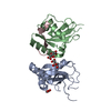
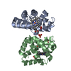
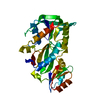
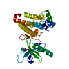
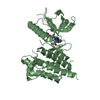
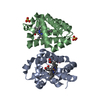
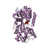
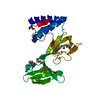
| Title | Crystal structure of human calmodulin-dependent protein kinase 1D (CAMK1D) bound to compound 18 (CS587) | ||||||
 Components Components | Calcium/calmodulin-dependent protein kinase type 1D | ||||||
 Keywords Keywords |  TRANSFERASE / Inhibitor / Complex / Kinase TRANSFERASE / Inhibitor / Complex / Kinase | ||||||
| Function / homology |  Function and homology information Function and homology informationregulation of granulocyte chemotaxis / positive regulation of CREB transcription factor activity / regulation of dendrite development / Ca2+/calmodulin-dependent protein kinase / positive regulation of neutrophil chemotaxis / calmodulin-dependent protein kinase activity / positive regulation of respiratory burst / positive regulation of phagocytosis / positive regulation of neuron projection development / nervous system development ...regulation of granulocyte chemotaxis / positive regulation of CREB transcription factor activity / regulation of dendrite development / Ca2+/calmodulin-dependent protein kinase / positive regulation of neutrophil chemotaxis / calmodulin-dependent protein kinase activity / positive regulation of respiratory burst / positive regulation of phagocytosis / positive regulation of neuron projection development / nervous system development / calmodulin binding / inflammatory response / positive regulation of apoptotic process / phosphorylation / protein serine kinase activity / negative regulation of apoptotic process / ATP binding / nucleus / cytoplasmSimilarity search - Function | ||||||
| Biological species |  Homo sapiens (human) Homo sapiens (human) | ||||||
| Method | X-RAY DIFFRACTION / SYNCHROTRON / MOLECULAR REPLACEMENT / Resolution: 1.484 Å | ||||||
 Authors Authors | Kraemer, A. / Sorrell, F. / Butterworth, S. / Edwards, A.M. / Arrowsmith, C.H. / Bountra, C. / Knapp, S. / Structural Genomics Consortium (SGC) | ||||||
| Funding support |  United Kingdom, 1items United Kingdom, 1items
| ||||||
 Citation Citation | Journal: J.Med.Chem. / Year: 2020 Title: Discovery of Highly Selective Inhibitors of Calmodulin-Dependent Kinases That Restore Insulin Sensitivity in the Diet-Induced Obesityin VivoMouse Model. Authors: Fromont, C. / Atzori, A. / Kaur, D. / Hashmi, L. / Greco, G. / Cabanillas, A. / Nguyen, H.V. / Jones, D.H. / Garzon, M. / Varela, A. / Stevenson, B. / Iacobini, G.P. / Lenoir, M. / Rajesh, S. ...Authors: Fromont, C. / Atzori, A. / Kaur, D. / Hashmi, L. / Greco, G. / Cabanillas, A. / Nguyen, H.V. / Jones, D.H. / Garzon, M. / Varela, A. / Stevenson, B. / Iacobini, G.P. / Lenoir, M. / Rajesh, S. / Box, C. / Kumar, J. / Grant, P. / Novitskaya, V. / Morgan, J. / Sorrell, F.J. / Redondo, C. / Kramer, A. / Harris, C.J. / Leighton, B. / Vickers, S.P. / Cheetham, S.C. / Kenyon, C. / Grabowska, A.M. / Overduin, M. / Berditchevski, F. / Weston, C.J. / Knapp, S. / Fischer, P.M. / Butterworth, S. | ||||||
| History |
|
- Structure visualization
| Structure viewer | Molecule: MolmilJmol/JSmol |
|---|
- Downloads & links
-Download
| PDBx/mmCIF format | 6t29.cif.gz | 130.9 KB | Display | PDBx/mmCIF format |
|---|---|---|---|---|
| PDB format | pdb6t29.ent.gz | Display | PDB format | |
| PDBx/mmJSON format | 6t29.json.gz | Tree view | PDBx/mmJSON format | |
| Others |  Other downloads Other downloads |
-Validation report
| Arichive directory | https://data.pdbj.org/pub/pdb/validation_reports/t2/6t29ftp://data.pdbj.org/pub/pdb/validation_reports/t2/6t29 | HTTPS FTP |
|---|
-Related structure data
| Related structure data |  6t28C  2jc6S S: Starting model for refinement C: citing same article ( |
|---|---|
| Similar structure data |


-Links
 PDBj
PDBj
- Assembly
| Deposited unit | 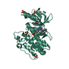
| |||||||||
|---|---|---|---|---|---|---|---|---|---|---|
| 1 |
| |||||||||
| Unit cell |
| |||||||||
| Components on special symmetry positions |
|
-Components
| #1: Protein | Mass: 42972.801 Da / Num. of mol.: 1 Source method: isolated from a genetically manipulated source Source: (gene. exp.) Homo sapiens (human) / Gene: CAMK1D, CAMKID / Production host:  Escherichia coli (E. coli) Escherichia coli (E. coli)References: UniProt: Q8IU85, Ca2+/calmodulin-dependent protein kinase | ||||||||
|---|---|---|---|---|---|---|---|---|---|
| #2: Chemical | ChemComp-SO4 / Sulfate  Mass: 96.063 Da / Num. of mol.: 4 / Source method: obtained synthetically / Formula: SO4 Mass: 96.063 Da / Num. of mol.: 4 / Source method: obtained synthetically / Formula: SO4#3: Chemical | ChemComp-EDO / Ethylene glycol  Mass: 62.068 Da / Num. of mol.: 5 / Source method: obtained synthetically / Formula: C2H6O2 Mass: 62.068 Da / Num. of mol.: 5 / Source method: obtained synthetically / Formula: C2H6O2#4: Chemical | ChemComp-M8Z / |   Mass: 446.548 Da / Num. of mol.: 1 / Source method: obtained synthetically / Formula: C24H30N8O / Feature type: SUBJECT OF INVESTIGATION Mass: 446.548 Da / Num. of mol.: 1 / Source method: obtained synthetically / Formula: C24H30N8O / Feature type: SUBJECT OF INVESTIGATION#5: Water | ChemComp-HOH / | Water Mass: 18.015 Da / Num. of mol.: 197 / Source method: isolated from a natural source / Formula: H2O Mass: 18.015 Da / Num. of mol.: 197 / Source method: isolated from a natural source / Formula: H2OHas ligand of interest | Y | |
-Experimental details
-Experiment
| Experiment | Method: X-RAY DIFFRACTION / Number of used crystals: 1 |
|---|
- Sample preparation
| Crystal grow | Temperature: 293 K / Method: vapor diffusion, sitting drop / pH: 5.9 / Details: 0,1M NaCit, pH 5.9 2,05M AmmSO4 0,1M Na/K tartrate |
|---|
-Data collection
| Diffraction | Mean temperature: 100 K / Serial crystal experiment: N |
|---|---|
| Diffraction source | Source: SYNCHROTRON / Site: PETRA III, EMBL c/o DESY  / Beamline: P13 (MX1) / Wavelength: 0.95372 Å / Beamline: P13 (MX1) / Wavelength: 0.95372 Å |
| Detector | Type: DECTRIS PILATUS 6M / Detector: PIXEL / Date: Sep 24, 2017 |
| Radiation | Protocol: SINGLE WAVELENGTH / Monochromatic (M) / Laue (L): M / Scattering type: x-ray |
| Radiation wavelength | Wavelength: 0.95372 Å / Relative weight: 1 |
| Reflection | Resolution: 1.48→55.45 Å / Num. obs: 44955 / % possible obs: 98.7 % / Redundancy: 6.3 % / CC1/2: 0.999 / Rpim(I) all: 0.026 / Net I/σ(I): 14.8 |
| Reflection shell | Resolution: 1.48→1.51 Å / Redundancy: 6.6 % / Mean I/σ(I) obs: 4.2 / Num. unique obs: 14295 / CC1/2: 0.883 / Rpim(I) all: 0.155 / % possible all: 98.3 |
- Processing
| Software |
| |||||||||||||||||||||||||||||||||||||||||||||||||||||||||||||||||||||||||||||||||||||||||||||||||||||||||||||||||||||||||||||||||||||||||||||||||||||||||||
|---|---|---|---|---|---|---|---|---|---|---|---|---|---|---|---|---|---|---|---|---|---|---|---|---|---|---|---|---|---|---|---|---|---|---|---|---|---|---|---|---|---|---|---|---|---|---|---|---|---|---|---|---|---|---|---|---|---|---|---|---|---|---|---|---|---|---|---|---|---|---|---|---|---|---|---|---|---|---|---|---|---|---|---|---|---|---|---|---|---|---|---|---|---|---|---|---|---|---|---|---|---|---|---|---|---|---|---|---|---|---|---|---|---|---|---|---|---|---|---|---|---|---|---|---|---|---|---|---|---|---|---|---|---|---|---|---|---|---|---|---|---|---|---|---|---|---|---|---|---|---|---|---|---|---|---|---|
| Refinement | Method to determine structure: MOLECULAR REPLACEMENT Starting model: 2jc6 Resolution: 1.484→55.448 Å / Cor.coef. Fo:Fc: 0.972 / Cor.coef. Fo:Fc free: 0.963 / SU B: 2.14 / SU ML: 0.036 / Cross valid method: FREE R-VALUE / ESU R: 0.072 / ESU R Free: 0.064 Details: Hydrogens have been added in their riding positions
| |||||||||||||||||||||||||||||||||||||||||||||||||||||||||||||||||||||||||||||||||||||||||||||||||||||||||||||||||||||||||||||||||||||||||||||||||||||||||||
| Solvent computation | Ion probe radii: 0.8 Å / Shrinkage radii: 0.8 Å / VDW probe radii: 1.2 Å | |||||||||||||||||||||||||||||||||||||||||||||||||||||||||||||||||||||||||||||||||||||||||||||||||||||||||||||||||||||||||||||||||||||||||||||||||||||||||||
| Displacement parameters | Biso mean: 17.818 Å2
| |||||||||||||||||||||||||||||||||||||||||||||||||||||||||||||||||||||||||||||||||||||||||||||||||||||||||||||||||||||||||||||||||||||||||||||||||||||||||||
| Refinement step | Cycle: LAST / Resolution: 1.484→55.448 Å
| |||||||||||||||||||||||||||||||||||||||||||||||||||||||||||||||||||||||||||||||||||||||||||||||||||||||||||||||||||||||||||||||||||||||||||||||||||||||||||
| Refine LS restraints |
| |||||||||||||||||||||||||||||||||||||||||||||||||||||||||||||||||||||||||||||||||||||||||||||||||||||||||||||||||||||||||||||||||||||||||||||||||||||||||||
| LS refinement shell |
|
