 Movie
Movie Controller


+ Open data

- Basic information
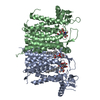
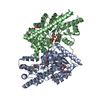
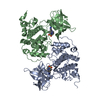
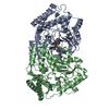
| Entry | Database: PDB / ID: 6sz6 | |||||||||
|---|---|---|---|---|---|---|---|---|---|---|
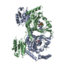
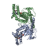
| Title | Chaetomium thermophilum beta-glucosidase | |||||||||
 Components Components | Beta-glucosidase | |||||||||
 Keywords Keywords | HYDROLASE / Cellulose degradation / thermophilic fungus / biofuel / glucosidase | |||||||||
| Function / homology |  Function and homology information Function and homology informationpolysaccharide catabolic process / hydrolase activity, hydrolyzing O-glycosyl compounds Similarity search - Function | |||||||||
| Biological species |  Chaetomium thermophilum (fungus) Chaetomium thermophilum (fungus) | |||||||||
| Method | X-RAY DIFFRACTION / SYNCHROTRON / MOLECULAR REPLACEMENT / Resolution: 2.988 Å | |||||||||
 Authors Authors | Mohsin, I. / Poudel, N. / Papageorgiou, A.C. | |||||||||
 Citation Citation | Journal: Int J Mol Sci / Year: 2019 Title: Crystal Structure of a GH3 beta-Glucosidase from the Thermophilic Fungus Chaetomium thermophilum . Authors: Mohsin, I. / Poudel, N. / Li, D.C. / Papageorgiou, A.C. | |||||||||
| History |
|
- Structure visualization
| Structure viewer | Molecule: MolmilJmol/JSmol |
|---|
- Downloads & links
-Download
| PDBx/mmCIF format | 6sz6.cif.gz | 354.2 KB | Display | PDBx/mmCIF format |
|---|---|---|---|---|
| PDB format | pdb6sz6.ent.gz | 286.9 KB | Display | PDB format |
| PDBx/mmJSON format | 6sz6.json.gz | Tree view | PDBx/mmJSON format | |
| Others |  Other downloads Other downloads |
-Validation report
| Arichive directory | https://data.pdbj.org/pub/pdb/validation_reports/sz/6sz6ftp://data.pdbj.org/pub/pdb/validation_reports/sz/6sz6 | HTTPS FTP |
|---|
-Related structure data
| Related structure data |  4iifS S: Starting model for refinement |
|---|---|
| Similar structure data |


-Links
 PDBj
PDBj


- Assembly
| Deposited unit | 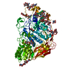
| ||||||||
|---|---|---|---|---|---|---|---|---|---|
| 1 |
| ||||||||
| Unit cell |
|
-Components
-Protein / Non-polymers , 2 types, 7 molecules A
| #10: Water | ChemComp-HOH / WaterMass: 18.015 Da / Num. of mol.: 6 / Source method: isolated from a natural source / Formula: H2O |
|---|---|
| #1: Protein | Mass: 91711.250 Da / Num. of mol.: 1 Source method: isolated from a genetically manipulated source Details: The first 16 correspond to the signal peptide. Residues 17-31 are not visible in the electron density map. Source: (gene. exp.) Chaetomium thermophilum (fungus) / Production host: Komagataella phaffii GS115 (fungus) / References: UniProt: A6YRT4 |
-Sugars , 8 types, 15 molecules 


| #2: Polysaccharide | / Mass: 586.542 Da / Num. of mol.: 3 Source method: isolated from a genetically manipulated source #3: Polysaccharide | / Mass: 424.401 Da / Num. of mol.: 2Source method: isolated from a genetically manipulated source #4: Polysaccharide | alpha-D-mannopyranose-(1-3)-beta-D-mannopyranose-(1-4)-2-acetamido-2-deoxy-beta-D-glucopyranose-(1- ...alpha-D-mannopyranose-(1-3)-beta-D-mannopyranose-(1-4)-2-acetamido-2-deoxy-beta-D-glucopyranose-(1-4)-2-acetamido-2-deoxy-beta-D-glucopyranose | / Mass: 748.682 Da / Num. of mol.: 1Source method: isolated from a genetically manipulated source #5: Polysaccharide | alpha-D-mannopyranose-(1-2)-alpha-D-mannopyranose-(1-6)-alpha-D-mannopyranose | / Mass: 504.438 Da / Num. of mol.: 1 / Source method: isolated from a natural source#6: Sugar | ChemComp-MAN / | Mannose Type: D-saccharide, alpha linking / Mass: 180.156 Da / Num. of mol.: 1 / Source method: isolated from a natural source / Formula: C6H12O6 Type: D-saccharide, alpha linking / Mass: 180.156 Da / Num. of mol.: 1 / Source method: isolated from a natural source / Formula: C6H12O6#7: Sugar | ChemComp-NAG / N-Acetylglucosamine Type: D-saccharide, beta linking / Mass: 221.208 Da / Num. of mol.: 4 Type: D-saccharide, beta linking / Mass: 221.208 Da / Num. of mol.: 4Source method: isolated from a genetically manipulated source Formula: C8H15NO6 #8: Sugar | Mannose Type: D-saccharide, beta linking / Mass: 180.156 Da / Num. of mol.: 2 Type: D-saccharide, beta linking / Mass: 180.156 Da / Num. of mol.: 2Source method: isolated from a genetically manipulated source Formula: C6H12O6 #9: Sugar | ChemComp-BGC / | Glucose Type: D-saccharide, beta linking / Mass: 180.156 Da / Num. of mol.: 1 Type: D-saccharide, beta linking / Mass: 180.156 Da / Num. of mol.: 1Source method: isolated from a genetically manipulated source Formula: C6H12O6 / Feature type: SUBJECT OF INVESTIGATION |
|---|
-Details
| Has ligand of interest | Y |
|---|
-Experimental details
-Experiment
| Experiment | Method: X-RAY DIFFRACTION / Number of used crystals: 1 |
|---|
- Sample preparation
| Crystal | Density Matthews: 5.4 Å3/Da / Density % sol: 77.06 % |
|---|---|
| Crystal grow | Temperature: 289 K / Method: vapor diffusion, hanging drop / pH: 7.6 / Details: 35-45 % v/v MPD, HEPES 0.1 M, pH 7.6 |
-Data collection
| Diffraction | Mean temperature: 100 K / Serial crystal experiment: N |
|---|---|
| Diffraction source | Source: SYNCHROTRON / Site: EMBL/DESY, HAMBURG  / Beamline: X13 / Wavelength: 0.8123 Å / Beamline: X13 / Wavelength: 0.8123 Å |
| Detector | Type: MAR CCD 165 mm / Detector: CCD / Date: May 6, 2011 |
| Radiation | Protocol: SINGLE WAVELENGTH / Monochromatic (M) / Laue (L): M / Scattering type: x-ray |
| Radiation wavelength | Wavelength: 0.8123 Å / Relative weight: 1 |
| Reflection | Resolution: 2.988→20 Å / Num. obs: 41052 / % possible obs: 99.3 % / Redundancy: 5.4 % / Biso Wilson estimate: 46.2 Å2 / CC1/2: 0.978 / Rpim(I) all: 0.122 / Rrim(I) all: 0.287 / Net I/σ(I): 7.8 |
| Reflection shell | Resolution: 2.99→3.11 Å / Redundancy: 5.1 % / Mean I/σ(I) obs: 1.4 / Num. unique obs: 4513 / CC1/2: 0.492 / Rpim(I) all: 0.846 / % possible all: 98.6 |
- Processing
| Software |
| ||||||||||||||||||||||||||||||||||||||||||||||||||||||||||||||||||||||||||||||||||||||||||||||||
|---|---|---|---|---|---|---|---|---|---|---|---|---|---|---|---|---|---|---|---|---|---|---|---|---|---|---|---|---|---|---|---|---|---|---|---|---|---|---|---|---|---|---|---|---|---|---|---|---|---|---|---|---|---|---|---|---|---|---|---|---|---|---|---|---|---|---|---|---|---|---|---|---|---|---|---|---|---|---|---|---|---|---|---|---|---|---|---|---|---|---|---|---|---|---|---|---|---|
| Refinement | Method to determine structure: MOLECULAR REPLACEMENT Starting model: 4iif Resolution: 2.988→19.831 Å / SU ML: 0.43 / Cross valid method: THROUGHOUT / σ(F): 1.33 / Phase error: 25.78 / Stereochemistry target values: ML
| ||||||||||||||||||||||||||||||||||||||||||||||||||||||||||||||||||||||||||||||||||||||||||||||||
| Solvent computation | Shrinkage radii: 0.9 Å / VDW probe radii: 1.11 Å / Solvent model: FLAT BULK SOLVENT MODEL | ||||||||||||||||||||||||||||||||||||||||||||||||||||||||||||||||||||||||||||||||||||||||||||||||
| Displacement parameters | Biso max: 146.86 Å2 / Biso mean: 51.5656 Å2 / Biso min: 24.13 Å2 | ||||||||||||||||||||||||||||||||||||||||||||||||||||||||||||||||||||||||||||||||||||||||||||||||
| Refinement step | Cycle: final / Resolution: 2.988→19.831 Å
| ||||||||||||||||||||||||||||||||||||||||||||||||||||||||||||||||||||||||||||||||||||||||||||||||
| LS refinement shell | Refine-ID: X-RAY DIFFRACTION / Rfactor Rfree error: 0
| ||||||||||||||||||||||||||||||||||||||||||||||||||||||||||||||||||||||||||||||||||||||||||||||||
| Refinement TLS params. | Method: refined / Origin x: -35.731 Å / Origin y: 4.0951 Å / Origin z: -42.264 Å
| ||||||||||||||||||||||||||||||||||||||||||||||||||||||||||||||||||||||||||||||||||||||||||||||||
| Refinement TLS group |
|
