 Movie
Movie Controller

[English] 日本語
 Yorodumi
- PDB-6swr: Crystal structure of the lysosomal potassium channel MtTMEM175 T3... -
+ Open data

- Basic information
| Entry | Database: PDB / ID: 6swr | |||||||||
|---|---|---|---|---|---|---|---|---|---|---|
| Title | Crystal structure of the lysosomal potassium channel MtTMEM175 T38A mutant soaked with zinc | |||||||||
 Components Components |
| |||||||||
 Keywords Keywords |  TRANSPORT PROTEIN / lysosome / TMEM175 / potassium channel / Parkinson disease TRANSPORT PROTEIN / lysosome / TMEM175 / potassium channel / Parkinson disease | |||||||||
| Function / homology |  Function and homology informationproton channel activity / potassium ion leak channel activity / detection of maltose stimulus / maltose transport complex / maltose binding / maltose transport / maltodextrin transmembrane transport / carbohydrate transport / carbohydrate transmembrane transporter activity / ATP-binding cassette (ABC) transporter complex, substrate-binding subunit-containing ...proton channel activity / potassium ion leak channel activity / detection of maltose stimulus / maltose transport complex / maltose binding / maltose transport / maltodextrin transmembrane transport / carbohydrate transport / carbohydrate transmembrane transporter activity / ATP-binding cassette (ABC) transporter complex, substrate-binding subunit-containing / potassium ion transmembrane transport / ATP-binding cassette (ABC) transporter complex / cell chemotaxis / outer membrane-bounded periplasmic space / protein homotetramerization / periplasmic space / DNA damage response / membrane / plasma membrane Function and homology informationproton channel activity / potassium ion leak channel activity / detection of maltose stimulus / maltose transport complex / maltose binding / maltose transport / maltodextrin transmembrane transport / carbohydrate transport / carbohydrate transmembrane transporter activity / ATP-binding cassette (ABC) transporter complex, substrate-binding subunit-containing ...proton channel activity / potassium ion leak channel activity / detection of maltose stimulus / maltose transport complex / maltose binding / maltose transport / maltodextrin transmembrane transport / carbohydrate transport / carbohydrate transmembrane transporter activity / ATP-binding cassette (ABC) transporter complex, substrate-binding subunit-containing / potassium ion transmembrane transport / ATP-binding cassette (ABC) transporter complex / cell chemotaxis / outer membrane-bounded periplasmic space / protein homotetramerization / periplasmic space / DNA damage response / membrane / plasma membraneSimilarity search - Function | |||||||||
| Biological species |  Lama glama (llama) Lama glama (llama) Escherichia coli (E. coli)Marivirga tractuosa Escherichia coli (E. coli)Marivirga tractuosa | |||||||||
| Method | X-RAY DIFFRACTION / SYNCHROTRON / MOLECULAR REPLACEMENT / Resolution: 3.2 Å | |||||||||
 Authors Authors | Brunner, J.D. / Jakob, R.P. / Schulze, T. / Neldner, Y. / Moroni, A. / Thiel, G. / Maier, T. / Schenck, S. | |||||||||
 Citation Citation | Journal: Elife / Year: 2020 Title: Structural basis for ion selectivity in TMEM175 K + channels. Authors: Brunner, J.D. / Jakob, R.P. / Schulze, T. / Neldner, Y. / Moroni, A. / Thiel, G. / Maier, T. / Schenck, S. | |||||||||
| History |
|
- Structure visualization
| Structure viewer | Molecule: MolmilJmol/JSmol |
|---|
- Downloads & links
-Download
| PDBx/mmCIF format | 6swr.cif.gz | 1.1 MB | Display | PDBx/mmCIF format |
|---|---|---|---|---|
| PDB format | pdb6swr.ent.gz | 919.1 KB | Display | PDB format |
| PDBx/mmJSON format | 6swr.json.gz | Tree view | PDBx/mmJSON format | |
| Others |  Other downloads Other downloads |
-Validation report
| Arichive directory | https://data.pdbj.org/pub/pdb/validation_reports/sw/6swrftp://data.pdbj.org/pub/pdb/validation_reports/sw/6swr | HTTPS FTP |
|---|
-Related structure data
| Related structure data | 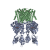 6hd8C 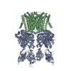 6hd9C  6hdaC 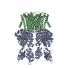 6hdbC  6hdcC  1anfS 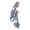 5jqhS C: citing same article ( S: Starting model for refinement |
|---|---|
| Similar structure data |


-Links
 PDBj
PDBj


- Assembly
| Deposited unit | 
| ||||||||
|---|---|---|---|---|---|---|---|---|---|
| 1 | 
| ||||||||
| Unit cell |
|
-Components
-Antibody / Protein , 2 types, 4 molecules ADBE
| #1: Antibody | Single-domain antibody / MMBP / Maltodextrin-binding protein / Maltose-binding protein / MBP Mass: 53267.004 Da / Num. of mol.: 2 Source method: isolated from a genetically manipulated source Source: (gene. exp.) Lama glama (llama), (gene. exp.) Escherichia coli (strain K12) (bacteria)Gene: malE, b4034, JW3994 / Plasmid: pBXNPHM3 / Strain: K12 / Production host: Escherichia coli MC1061 (bacteria) / References: UniProt: P0AEX9#2: Protein | Mass: 29450.943 Da / Num. of mol.: 2 Source method: isolated from a genetically manipulated source Source: (gene. exp.) Marivirga tractuosa (strain ATCC 23168 / DSM 4126 / NBRC 15989 / NCIMB 1408 / VKM B-1430 / H-43) (bacteria)Strain: ATCC 23168 / DSM 4126 / NBRC 15989 / NCIMB 1408 / VKM B-1430 / H-43 Gene: Ftrac_2467 / Production host: Escherichia coli MC1061 (bacteria) / References: UniProt: E4TN31 |
|---|
-Sugars , 2 types, 4 molecules 
| #3: Polysaccharide |   , Oligosaccharide / Class: Nutrient / Mass: 342.297 Da / Num. of mol.: 2 , Oligosaccharide / Class: Nutrient / Mass: 342.297 Da / Num. of mol.: 2Source method: isolated from a genetically manipulated source Details: oligosaccharide / References: alpha-maltose #4: Sugar |  Type: D-saccharide / Mass: 510.615 Da / Num. of mol.: 2 / Source method: obtained synthetically / Formula: C24H46O11 / Comment: detergent*YM Type: D-saccharide / Mass: 510.615 Da / Num. of mol.: 2 / Source method: obtained synthetically / Formula: C24H46O11 / Comment: detergent*YM |
|---|
-Non-polymers , 2 types, 32 molecules 

| #5: Chemical | ChemComp-K /  Mass: 39.098 Da / Num. of mol.: 1 / Source method: obtained synthetically / Formula: K / Feature type: SUBJECT OF INVESTIGATION Mass: 39.098 Da / Num. of mol.: 1 / Source method: obtained synthetically / Formula: K / Feature type: SUBJECT OF INVESTIGATION |
|---|---|
| #6: Water | ChemComp-HOH / WaterMass: 18.015 Da / Num. of mol.: 31 / Source method: isolated from a natural source / Formula: H2O |
-Details
| Has ligand of interest | Y |
|---|
-Experimental details
-Experiment
| Experiment | Method: X-RAY DIFFRACTION / Number of used crystals: 1 |
|---|
- Sample preparation
| Crystal | Density Matthews: 4.24 Å3/Da / Density % sol: 70.96 % |
|---|---|
| Crystal grow | Temperature: 293 K / Method: vapor diffusion, sitting drop Details: 100 mM Tris-HCl pH 8.5, 150 mM NaCl, 150 mM MgCl2 and 28-30 % PEG400 |
-Data collection
| Diffraction | Mean temperature: 100 K / Serial crystal experiment: N |
|---|---|
| Diffraction source | Source: SYNCHROTRON / Site: SLS  / Beamline: X06SA / Wavelength: 1.280961 Å / Beamline: X06SA / Wavelength: 1.280961 Å |
| Detector | Type: DECTRIS EIGER X 16M / Detector: PIXEL / Date: Feb 9, 2018 |
| Radiation | Protocol: SINGLE WAVELENGTH / Monochromatic (M) / Laue (L): M / Scattering type: x-ray |
| Radiation wavelength | Wavelength: 1.280961 Å / Relative weight: 1 |
| Reflection | Resolution: 3.2→49.72 Å / Num. obs: 43526 / % possible obs: 99.8 % / Redundancy: 51.8 % / Biso Wilson estimate: 76.29 Å2 / CC1/2: 0.993 / Net I/σ(I): 6.5 |
| Reflection shell | Resolution: 3.2→3.32 Å / Num. unique obs: 4510 / CC1/2: 0.663 |
- Processing
| Software |
| ||||||||||||||||
|---|---|---|---|---|---|---|---|---|---|---|---|---|---|---|---|---|---|
| Refinement | Method to determine structure: MOLECULAR REPLACEMENT Starting model: 1ANF, 5JQH Resolution: 3.2→49.72 Å / Cross valid method: FREE R-VALUE
| ||||||||||||||||
| Refinement step | Cycle: LAST / Resolution: 3.2→49.72 Å
|
