 Movie
Movie Controller

[English] 日本語
 Yorodumi
- PDB-6sua: Structure of the high affinity engineered lipocalin C1B12 in comp... -
+ Open data

- Basic information
| Entry | Database: PDB / ID: 6sua | ||||||
|---|---|---|---|---|---|---|---|
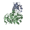
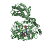
| Title | Structure of the high affinity engineered lipocalin C1B12 in complex with the mouse CD98 heavy chain ectodomain | ||||||
 Components Components |
| ||||||
 Keywords Keywords |  PROTEIN BINDING / Lipocalin / Lcn2 / Anticalin / lipocalin-based binding protein / CD98hc / 4F2hc / amino acid transport / presentation of CD98 light chain / interaction with integrin beta subunit / single-pass type II membrane protein PROTEIN BINDING / Lipocalin / Lcn2 / Anticalin / lipocalin-based binding protein / CD98hc / 4F2hc / amino acid transport / presentation of CD98 light chain / interaction with integrin beta subunit / single-pass type II membrane protein | ||||||
| Function / homology |  Function and homology information Function and homology informationAmino acid transport across the plasma membrane / Tryptophan catabolism / apical pole of neuron / aromatic amino acid transmembrane transporter activity / amino acid transport complex / : / L-leucine import across plasma membrane / L-alanine transmembrane transporter activity / L-alanine import across plasma membrane / phenylalanine transport ...Amino acid transport across the plasma membrane / Tryptophan catabolism / apical pole of neuron / aromatic amino acid transmembrane transporter activity / amino acid transport complex / : / L-leucine import across plasma membrane / L-alanine transmembrane transporter activity / L-alanine import across plasma membrane / phenylalanine transport / L-leucine transmembrane transporter activity / L-leucine transport / neutral amino acid transport / siderophore transport / Metal sequestration by antimicrobial proteins / neutral L-amino acid transmembrane transporter activity / iron ion sequestering activity / enterobactin binding / anchoring junction / response to exogenous dsRNA / tryptophan transport / basal plasma membrane / Iron uptake and transport / specific granule lumen / double-stranded RNA binding / melanosome / positive regulation of cold-induced thermogenesis / basolateral plasma membrane / Interleukin-4 and Interleukin-13 signaling / carbohydrate metabolic process / defense response to bacterium / symbiont entry into host cell / iron ion binding / apical plasma membrane / lysosomal membrane / innate immune response / neuronal cell body / apoptotic process / synapse / Neutrophil degranulation / cell surface / extracellular space / extracellular exosome / extracellular region / nucleoplasm / identical protein binding / plasma membrane / cytoplasmSimilarity search - Function | ||||||
| Biological species |  Homo sapiens (human) Homo sapiens (human) Mus musculus (house mouse) Mus musculus (house mouse) | ||||||
| Method | X-RAY DIFFRACTION / SYNCHROTRON / MOLECULAR REPLACEMENT / Resolution: 2.75 Å | ||||||
 Authors Authors | Schiefner, A. / Deuschle, F.-C. / Skerra, A. | ||||||
| Funding support |  Germany, 1items Germany, 1items
| ||||||
 Citation Citation | Journal: Protein Sci. / Year: 2020 Title: Design of a surrogate Anticalin protein directed against CD98hc for preclinical studies in mice. Authors: Deuschle, F.C. / Schiefner, A. / Brandt, C. / Skerra, A. | ||||||
| History |
|
- Structure visualization
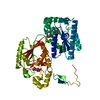
| Structure viewer | Molecule: MolmilJmol/JSmol |
|---|
- Downloads & links
-Download
| PDBx/mmCIF format | 6sua.cif.gz | 248 KB | Display | PDBx/mmCIF format |
|---|---|---|---|---|
| PDB format | pdb6sua.ent.gz | 198 KB | Display | PDB format |
| PDBx/mmJSON format | 6sua.json.gz | Tree view | PDBx/mmJSON format | |
| Others |  Other downloads Other downloads |
-Validation report
| Arichive directory | https://data.pdbj.org/pub/pdb/validation_reports/su/6suaftp://data.pdbj.org/pub/pdb/validation_reports/su/6sua | HTTPS FTP |
|---|
-Related structure data


-Links
 PDBj
PDBj


- Assembly
| Deposited unit | 
| ||||||||||||||||||||||||||||||||||||||||||||||||||||||||||||||||||||
|---|---|---|---|---|---|---|---|---|---|---|---|---|---|---|---|---|---|---|---|---|---|---|---|---|---|---|---|---|---|---|---|---|---|---|---|---|---|---|---|---|---|---|---|---|---|---|---|---|---|---|---|---|---|---|---|---|---|---|---|---|---|---|---|---|---|---|---|---|---|
| 1 | 
| ||||||||||||||||||||||||||||||||||||||||||||||||||||||||||||||||||||
| 2 | 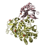
| ||||||||||||||||||||||||||||||||||||||||||||||||||||||||||||||||||||
| Unit cell |
| ||||||||||||||||||||||||||||||||||||||||||||||||||||||||||||||||||||
| Noncrystallographic symmetry (NCS) | NCS domain:
NCS domain segments: Component-ID: 0 / Refine code: 0
NCS ensembles :
|
-Components
| #1: Protein | Mass: 21462.252 Da / Num. of mol.: 2 Mutation: Q28H, A40G, I41N, Q49L, S68D, R72I, K73D, D77H, W79L, R81N, C87S, N96S, Y100T, P101L, L103S, Y106R, K125W, S127H, Y132W, K134Y Source method: isolated from a genetically manipulated source Source: (gene. exp.) Homo sapiens (human) / Gene: LCN2, HNL, NGAL / Plasmid: pNGAL118 / Production host:  Escherichia coli (E. coli) / Strain (production host): JM83 / References: UniProt: P80188 Escherichia coli (E. coli) / Strain (production host): JM83 / References: UniProt: P80188#2: Protein | / 4F2hc / Solute carrier family 3 member 2Mass: 48366.570 Da / Num. of mol.: 2 Source method: isolated from a genetically manipulated source Source: (gene. exp.) Mus musculus (house mouse) / Gene: Slc3a2, Mdu1 / Plasmid: pASK-IBA5(+) / Production host: Escherichia coli BL21 (bacteria) / References: UniProt: P10852#3: Chemical | ChemComp-SO4 / Sulfate  Mass: 96.063 Da / Num. of mol.: 12 / Source method: obtained synthetically / Formula: SO4 Mass: 96.063 Da / Num. of mol.: 12 / Source method: obtained synthetically / Formula: SO4#4: Water | ChemComp-HOH / | Water Mass: 18.015 Da / Num. of mol.: 146 / Source method: isolated from a natural source / Formula: H2O Mass: 18.015 Da / Num. of mol.: 146 / Source method: isolated from a natural source / Formula: H2OHas ligand of interest | N | |
|---|
-Experimental details
-Experiment
| Experiment | Method: X-RAY DIFFRACTION / Number of used crystals: 1 |
|---|
- Sample preparation
| Crystal | Density Matthews: 2.71 Å3/Da / Density % sol: 54.65 % |
|---|---|
| Crystal grow | Temperature: 293 K / Method: vapor diffusion, hanging drop / pH: 7.5 / Details: 12 %(w/v) PEG8000, 0.2 M Li2SO4 |
-Data collection
| Diffraction | Mean temperature: 100 K / Serial crystal experiment: N | ||||||||||||||||||||||||||||||||||||||||||||||||||||||||||||||||||||||||||||||||||||||||||||||||||||
|---|---|---|---|---|---|---|---|---|---|---|---|---|---|---|---|---|---|---|---|---|---|---|---|---|---|---|---|---|---|---|---|---|---|---|---|---|---|---|---|---|---|---|---|---|---|---|---|---|---|---|---|---|---|---|---|---|---|---|---|---|---|---|---|---|---|---|---|---|---|---|---|---|---|---|---|---|---|---|---|---|---|---|---|---|---|---|---|---|---|---|---|---|---|---|---|---|---|---|---|---|---|
| Diffraction source | Source: SYNCHROTRON / Site: BESSY / Beamline: 14.2 / Wavelength: 0.9184 Å | ||||||||||||||||||||||||||||||||||||||||||||||||||||||||||||||||||||||||||||||||||||||||||||||||||||
| Detector | Type: DECTRIS PILATUS3 2M / Detector: PIXEL / Date: Mar 22, 2018 | ||||||||||||||||||||||||||||||||||||||||||||||||||||||||||||||||||||||||||||||||||||||||||||||||||||
| Radiation | Protocol: SINGLE WAVELENGTH / Monochromatic (M) / Laue (L): M / Scattering type: x-ray | ||||||||||||||||||||||||||||||||||||||||||||||||||||||||||||||||||||||||||||||||||||||||||||||||||||
| Radiation wavelength | Wavelength: 0.9184 Å / Relative weight: 1 | ||||||||||||||||||||||||||||||||||||||||||||||||||||||||||||||||||||||||||||||||||||||||||||||||||||
| Reflection | Resolution: 2.75→34.71 Å / Num. obs: 39856 / % possible obs: 99.1 % / Redundancy: 13.198 % / Biso Wilson estimate: 42.73 Å2 / CC1/2: 0.996 / Rmerge(I) obs: 0.197 / Rrim(I) all: 0.205 / Χ2: 0.944 / Net I/σ(I): 15.36 / Num. measured all: 526014 / Scaling rejects: 244 | ||||||||||||||||||||||||||||||||||||||||||||||||||||||||||||||||||||||||||||||||||||||||||||||||||||
| Reflection shell | Diffraction-ID: 1
|
- Processing
| Software |
| ||||||||||||||||||||||||||||||||||||||||||||||||||||||||||||
|---|---|---|---|---|---|---|---|---|---|---|---|---|---|---|---|---|---|---|---|---|---|---|---|---|---|---|---|---|---|---|---|---|---|---|---|---|---|---|---|---|---|---|---|---|---|---|---|---|---|---|---|---|---|---|---|---|---|---|---|---|---|
| Refinement | Method to determine structure: MOLECULAR REPLACEMENT Starting model: 6I9Q, 6S8V Resolution: 2.75→34.71 Å / Cor.coef. Fo:Fc: 0.919 / Cor.coef. Fo:Fc free: 0.874 / WRfactor Rfree: 0.2251 / WRfactor Rwork: 0.1825 / FOM work R set: 0.7783 / SU B: 16.437 / SU ML: 0.314 / SU Rfree: 0.3831 / Cross valid method: THROUGHOUT / σ(F): 0 / ESU R Free: 0.383 / Stereochemistry target values: MAXIMUM LIKELIHOOD Details: HYDROGENS HAVE BEEN ADDED IN THE RIDING POSITIONS U VALUES : REFINED INDIVIDUALLY
| ||||||||||||||||||||||||||||||||||||||||||||||||||||||||||||
| Solvent computation | Ion probe radii: 0.8 Å / Shrinkage radii: 0.8 Å / VDW probe radii: 1.2 Å / Solvent model: MASK | ||||||||||||||||||||||||||||||||||||||||||||||||||||||||||||
| Displacement parameters | Biso max: 145.8 Å2 / Biso mean: 43.758 Å2 / Biso min: 12.68 Å2
| ||||||||||||||||||||||||||||||||||||||||||||||||||||||||||||
| Refinement step | Cycle: final / Resolution: 2.75→34.71 Å
| ||||||||||||||||||||||||||||||||||||||||||||||||||||||||||||
| Refine LS restraints |
| ||||||||||||||||||||||||||||||||||||||||||||||||||||||||||||
| Refine LS restraints NCS | Refine-ID: X-RAY DIFFRACTION / Type: interatomic distance / Weight position: 0.05
| ||||||||||||||||||||||||||||||||||||||||||||||||||||||||||||
| LS refinement shell | Resolution: 2.75→2.821 Å / Rfactor Rfree error: 0 / Total num. of bins used: 20
|
