 Movie
Movie Controller

[English] 日本語
 Yorodumi
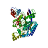
- PDB-6ssu: Crystal structure of Human Microsomal Glutathione S-Transferase 2... -
+ Open data

- Basic information
| Entry | Database: PDB / ID: 6ssu | ||||||
|---|---|---|---|---|---|---|---|
| Title | Crystal structure of Human Microsomal Glutathione S-Transferase 2 in complex with co-substrate Glutathione | ||||||
 Components Components | Microsomal glutathione S-transferase 2 | ||||||
 Keywords Keywords |  TRANSFERASE / ER MEMBRANE PROTEIN / GLUTATHIONE TRANSFERASE / MAPEG / MGST2 / INTEGRAL MEMBRANE ENZYME TRANSFERASE / ER MEMBRANE PROTEIN / GLUTATHIONE TRANSFERASE / MAPEG / MGST2 / INTEGRAL MEMBRANE ENZYME | ||||||
| Function / homology |  Function and homology information Function and homology informationmembrane lipid catabolic process / leukotriene-C4 synthase / leukotriene-C4 synthase activity / glutathione biosynthetic process / Aflatoxin activation and detoxification / glutathione binding / Glutathione conjugation / leukotriene biosynthetic process / glutathione peroxidase activity / glutathione transferase ...membrane lipid catabolic process / leukotriene-C4 synthase / leukotriene-C4 synthase activity / glutathione biosynthetic process / Aflatoxin activation and detoxification / glutathione binding / Glutathione conjugation / leukotriene biosynthetic process / glutathione peroxidase activity / glutathione transferase / glutathione transferase activity / enzyme activator activity / Oxidoreductases; Acting on a peroxide as acceptor; Peroxidases / response to organonitrogen compound / lipid metabolic process / positive regulation of inflammatory response / nuclear envelope / response to lipopolysaccharide / intracellular membrane-bounded organelle / endoplasmic reticulum membrane / endoplasmic reticulum / membrane / identical protein binding / plasma membraneSimilarity search - Function | ||||||
| Biological species |  Homo sapiens (human) Homo sapiens (human) | ||||||
| Method | X-RAY DIFFRACTION / SYNCHROTRON / MOLECULAR REPLACEMENT / Resolution: 2.499 Å | ||||||
 Authors Authors | Thulasingam, M. / Haeggstrom, J.Z. | ||||||
| Funding support |  Sweden, 1items Sweden, 1items
| ||||||
 Citation Citation | Journal: Nat Commun / Year: 2021 Title: Crystal structures of human MGST2 reveal synchronized conformational changes regulating catalysis. Authors: Thulasingam, M. / Orellana, L. / Nji, E. / Ahmad, S. / Rinaldo-Matthis, A. / Haeggstrom, J.Z. | ||||||
| History |
|
- Structure visualization
| Structure viewer | Molecule: MolmilJmol/JSmol |
|---|
- Downloads & links
-Download
| PDBx/mmCIF format | 6ssu.cif.gz | 97.1 KB | Display | PDBx/mmCIF format |
|---|---|---|---|---|
| PDB format | pdb6ssu.ent.gz | 72.8 KB | Display | PDB format |
| PDBx/mmJSON format | 6ssu.json.gz | Tree view | PDBx/mmJSON format | |
| Others |  Other downloads Other downloads |
-Validation report
| Arichive directory | https://data.pdbj.org/pub/pdb/validation_reports/ss/6ssuftp://data.pdbj.org/pub/pdb/validation_reports/ss/6ssu | HTTPS FTP |
|---|
-Related structure data
| Related structure data | 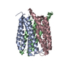 6ssrSC 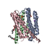 6sssC 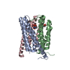 6sswC S: Starting model for refinement C: citing same article ( |
|---|---|
| Similar structure data |


-Links
 PDBj
PDBj


- Assembly
| Deposited unit | 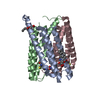
| ||||||||||||||||||||||||||||||||||||||||||||||||||||
|---|---|---|---|---|---|---|---|---|---|---|---|---|---|---|---|---|---|---|---|---|---|---|---|---|---|---|---|---|---|---|---|---|---|---|---|---|---|---|---|---|---|---|---|---|---|---|---|---|---|---|---|---|---|
| 1 |
| ||||||||||||||||||||||||||||||||||||||||||||||||||||
| Unit cell |
| ||||||||||||||||||||||||||||||||||||||||||||||||||||
| Noncrystallographic symmetry (NCS) | NCS domain:
NCS domain segments: Component-ID: 1 / Ens-ID: 1
|
-Components
| #1: Protein | Mass: 17465.410 Da / Num. of mol.: 3 Source method: isolated from a genetically manipulated source Source: (gene. exp.) Homo sapiens (human) / Gene: MGST2, GST2 / Production host:  Komagataella pastoris (fungus) / References: UniProt: Q99735, glutathione transferase Komagataella pastoris (fungus) / References: UniProt: Q99735, glutathione transferase#2: Chemical |   Mass: 356.540 Da / Num. of mol.: 2 / Source method: obtained synthetically / Formula: C21H40O4 Mass: 356.540 Da / Num. of mol.: 2 / Source method: obtained synthetically / Formula: C21H40O4#3: Chemical | Glutathione  Mass: 307.323 Da / Num. of mol.: 2 / Source method: obtained synthetically / Formula: C10H17N3O6S / Feature type: SUBJECT OF INVESTIGATION Mass: 307.323 Da / Num. of mol.: 2 / Source method: obtained synthetically / Formula: C10H17N3O6S / Feature type: SUBJECT OF INVESTIGATION#4: Chemical | ChemComp-NO3 / | Nitrate  Mass: 62.005 Da / Num. of mol.: 1 / Source method: obtained synthetically / Formula: NO3 Mass: 62.005 Da / Num. of mol.: 1 / Source method: obtained synthetically / Formula: NO3#5: Water | ChemComp-HOH / | Water Mass: 18.015 Da / Num. of mol.: 46 / Source method: isolated from a natural source / Formula: H2O Mass: 18.015 Da / Num. of mol.: 46 / Source method: isolated from a natural source / Formula: H2OHas ligand of interest | Y | |
|---|
-Experimental details
-Experiment
| Experiment | Method: X-RAY DIFFRACTION / Number of used crystals: 1 |
|---|
- Sample preparation
| Crystal | Density Matthews: 3.32 Å3/Da / Density % sol: 62.9 % |
|---|---|
| Crystal grow | Temperature: 298 K / Method: lipidic cubic phase / pH: 5.5 Details: 0.1M MES pH 5.5 0.4M Lithium citrate 0.1M Sodium nitrate 20% PEG 400 |
-Data collection
| Diffraction | Mean temperature: 100 K / Serial crystal experiment: N |
|---|---|
| Diffraction source | Source: SYNCHROTRON / Site: Diamond  / Beamline: I04-1 / Wavelength: 0.9282 Å / Beamline: I04-1 / Wavelength: 0.9282 Å |
| Detector | Type: DECTRIS PILATUS3 S 6M / Detector: PIXEL / Date: Apr 16, 2016 |
| Radiation | Protocol: SINGLE WAVELENGTH / Monochromatic (M) / Laue (L): M / Scattering type: x-ray |
| Radiation wavelength | Wavelength: 0.9282 Å / Relative weight: 1 |
| Reflection | Resolution: 2.49→46.28 Å / Num. obs: 21514 / % possible obs: 99.79 % / Redundancy: 7.5 % / Biso Wilson estimate: 58.65 Å2 / CC1/2: 0.999 / Net I/σ(I): 11.12 |
| Reflection shell | Resolution: 2.49→2.58 Å / Redundancy: 7.7 % / Mean I/σ(I) obs: 1.45 / Num. unique obs: 2106 / CC1/2: 0.605 / % possible all: 99.25 |
- Processing
| Software |
| ||||||||||||||||||||||||||||||||||||||||||||||||||||||
|---|---|---|---|---|---|---|---|---|---|---|---|---|---|---|---|---|---|---|---|---|---|---|---|---|---|---|---|---|---|---|---|---|---|---|---|---|---|---|---|---|---|---|---|---|---|---|---|---|---|---|---|---|---|---|---|
| Refinement | Method to determine structure: MOLECULAR REPLACEMENT Starting model: 6SSR Resolution: 2.499→46.28 Å / SU ML: 0.35 / Cross valid method: THROUGHOUT / σ(F): 1.35 / Phase error: 32.31 / Stereochemistry target values: ML
| ||||||||||||||||||||||||||||||||||||||||||||||||||||||
| Solvent computation | Shrinkage radii: 0.9 Å / VDW probe radii: 1.11 Å / Solvent model: FLAT BULK SOLVENT MODEL | ||||||||||||||||||||||||||||||||||||||||||||||||||||||
| Displacement parameters | Biso max: 127.18 Å2 / Biso mean: 59.8245 Å2 / Biso min: 32.7 Å2 | ||||||||||||||||||||||||||||||||||||||||||||||||||||||
| Refinement step | Cycle: final / Resolution: 2.499→46.28 Å
| ||||||||||||||||||||||||||||||||||||||||||||||||||||||
| Refine LS restraints |
| ||||||||||||||||||||||||||||||||||||||||||||||||||||||
| Refine LS restraints NCS |
| ||||||||||||||||||||||||||||||||||||||||||||||||||||||
| LS refinement shell | Refine-ID: X-RAY DIFFRACTION / Rfactor Rfree error: 0
|
