 Movie
Movie Controller

[English] 日本語
 Yorodumi
- PDB-6sr8: Crystal structure of glutathione transferase Omega 2C from Tramet... -
+ Open data

- Basic information
| Entry | Database: PDB / ID: 6sr8 | ||||||
|---|---|---|---|---|---|---|---|
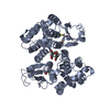
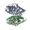
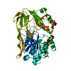
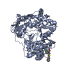
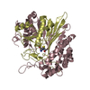
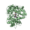
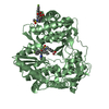
| Title | Crystal structure of glutathione transferase Omega 2C from Trametes versicolor | ||||||
 Components Components | Uncharacterized protein | ||||||
 Keywords Keywords |  TRANSFERASE / glutathione transferase TRANSFERASE / glutathione transferase | ||||||
| Function / homology |  Function and homology informationGlutathione S-transferase, N-terminal domain / Glutathione S-transferase, N-terminal domain / Glutathione S-transferase, C-terminal domain / Glutathione transferase family / Soluble glutathione S-transferase N-terminal domain profile. / Glutathione S-transferase, N-terminal / Glutathione S-transferase, C-terminal domain superfamily / Thioredoxin-like superfamily Function and homology informationGlutathione S-transferase, N-terminal domain / Glutathione S-transferase, N-terminal domain / Glutathione S-transferase, C-terminal domain / Glutathione transferase family / Soluble glutathione S-transferase N-terminal domain profile. / Glutathione S-transferase, N-terminal / Glutathione S-transferase, C-terminal domain superfamily / Thioredoxin-like superfamilySimilarity search - Domain/homology | ||||||
| Biological species |  Trametes versicolor (fungus) Trametes versicolor (fungus) | ||||||
| Method | X-RAY DIFFRACTION / SYNCHROTRON / MOLECULAR REPLACEMENT / Resolution: 1.94 Å | ||||||
 Authors Authors | Schwartz, M. / Favier, F. / Didierjean, C. | ||||||
| Funding support |  France, 1items France, 1items
| ||||||
 Citation Citation | Journal: Fungal Genet Biol. / Year: 2021 Title: Diversity of Omega Glutathione Transferases in mushroom-forming fungi revealed by phylogenetic, transcriptomic, biochemical and structural approaches. Authors: Perrot, T. / Schwartz, M. / Deroy, A. / Girardet, J.M. / Kohler, A. / Morel-Rouhier, M. / Favier, F. / Gelhaye, E. / Didierjean, C. | ||||||
| History |
|
- Structure visualization
| Structure viewer | Molecule: MolmilJmol/JSmol |
|---|
- Downloads & links
-Download
| PDBx/mmCIF format | 6sr8.cif.gz | 191.2 KB | Display | PDBx/mmCIF format |
|---|---|---|---|---|
| PDB format | pdb6sr8.ent.gz | 153.6 KB | Display | PDB format |
| PDBx/mmJSON format | 6sr8.json.gz | Tree view | PDBx/mmJSON format | |
| Others |  Other downloads Other downloads |
-Validation report
| Arichive directory | https://data.pdbj.org/pub/pdb/validation_reports/sr/6sr8ftp://data.pdbj.org/pub/pdb/validation_reports/sr/6sr8 | HTTPS FTP |
|---|
-Related structure data
| Related structure data |  6hjsSC  6sr9C  6sraC  6srbC S: Starting model for refinement C: citing same article ( |
|---|---|
| Similar structure data |


-Links
 PDBj
PDBj

- Assembly
| Deposited unit | 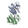
| ||||||||||||
|---|---|---|---|---|---|---|---|---|---|---|---|---|---|
| 1 | 
| ||||||||||||
| 2 | 
| ||||||||||||
| Unit cell |
| ||||||||||||
| Components on special symmetry positions |
|
-Components
| #1: Protein | Mass: 27428.176 Da / Num. of mol.: 2 Source method: isolated from a genetically manipulated source Source: (gene. exp.) Trametes versicolor (strain FP-101664) (fungus)Strain: FP-101664 / Gene: TRAVEDRAFT_67635 / Production host:  Escherichia coli (E. coli) / References: UniProt: R7S7J5 Escherichia coli (E. coli) / References: UniProt: R7S7J5#2: Chemical | Glutathione  Mass: 307.323 Da / Num. of mol.: 2 / Source method: obtained synthetically / Formula: C10H17N3O6S / Feature type: SUBJECT OF INVESTIGATION Mass: 307.323 Da / Num. of mol.: 2 / Source method: obtained synthetically / Formula: C10H17N3O6S / Feature type: SUBJECT OF INVESTIGATION#3: Water | ChemComp-HOH / | Water Mass: 18.015 Da / Num. of mol.: 386 / Source method: isolated from a natural source / Formula: H2O Mass: 18.015 Da / Num. of mol.: 386 / Source method: isolated from a natural source / Formula: H2OHas ligand of interest | Y | |
|---|
-Experimental details
-Experiment
| Experiment | Method: X-RAY DIFFRACTION / Number of used crystals: 1 |
|---|
- Sample preparation
| Crystal | Density Matthews: 2.77 Å3/Da / Density % sol: 55.64 % |
|---|---|
| Crystal grow | Temperature: 277 K / Method: microbatch Details: 20% (w/v) polyethylene glycol 4000, 10% (v/v) 2-Propanol and 0.1 M pH 7.5 HEPES |
-Data collection
| Diffraction | Mean temperature: 100 K / Serial crystal experiment: N |
|---|---|
| Diffraction source | Source: SYNCHROTRON / Site: ESRF / Beamline: BM30A / Wavelength: 0.979767 Å |
| Detector | Type: ADSC QUANTUM 315r / Detector: CCD / Date: Jun 29, 2015 |
| Radiation | Protocol: SINGLE WAVELENGTH / Monochromatic (M) / Laue (L): M / Scattering type: x-ray |
| Radiation wavelength | Wavelength: 0.979767 Å / Relative weight: 1 |
| Reflection | Resolution: 1.9→22.147 Å / Num. obs: 42824 / % possible obs: 97.1 % / Redundancy: 3.7 % / Biso Wilson estimate: 25.18 Å2 / CC1/2: 1 / Rmerge(I) obs: 0.055 / Rrim(I) all: 0.064 / Net I/σ(I): 13.33 |
| Reflection shell | Resolution: 1.9→1.95 Å / Rmerge(I) obs: 0.25 / Mean I/σ(I) obs: 5.28 / Num. unique obs: 3095 / CC1/2: 0.96 / Rrim(I) all: 0.3 |
- Processing
| Software |
| ||||||||||||||||||||||||||||||||||||||||||||||||||||||||||||||||||||||||||||||||||||||||||
|---|---|---|---|---|---|---|---|---|---|---|---|---|---|---|---|---|---|---|---|---|---|---|---|---|---|---|---|---|---|---|---|---|---|---|---|---|---|---|---|---|---|---|---|---|---|---|---|---|---|---|---|---|---|---|---|---|---|---|---|---|---|---|---|---|---|---|---|---|---|---|---|---|---|---|---|---|---|---|---|---|---|---|---|---|---|---|---|---|---|---|---|
| Refinement | Method to determine structure: MOLECULAR REPLACEMENT Starting model: 6HJS Resolution: 1.94→22.147 Å / SU ML: 0.22 / Cross valid method: FREE R-VALUE / σ(F): 1.34 / Phase error: 31.03
| ||||||||||||||||||||||||||||||||||||||||||||||||||||||||||||||||||||||||||||||||||||||||||
| Solvent computation | Shrinkage radii: 0.9 Å / VDW probe radii: 1.11 Å | ||||||||||||||||||||||||||||||||||||||||||||||||||||||||||||||||||||||||||||||||||||||||||
| Displacement parameters | Biso max: 105.19 Å2 / Biso mean: 39.0508 Å2 / Biso min: 16.19 Å2 | ||||||||||||||||||||||||||||||||||||||||||||||||||||||||||||||||||||||||||||||||||||||||||
| Refinement step | Cycle: final / Resolution: 1.94→22.147 Å
| ||||||||||||||||||||||||||||||||||||||||||||||||||||||||||||||||||||||||||||||||||||||||||
| Refine LS restraints |
| ||||||||||||||||||||||||||||||||||||||||||||||||||||||||||||||||||||||||||||||||||||||||||
| LS refinement shell | Refine-ID: X-RAY DIFFRACTION / Rfactor Rfree error: 0
|
