 Movie
Movie Controller


+ Open data

- Basic information
| Entry | Database: PDB / ID: 6si6 | |||||||||
|---|---|---|---|---|---|---|---|---|---|---|
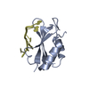
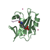
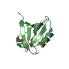
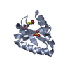
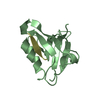
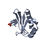
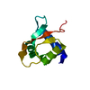
| Title | N-terminal domain of Drosophila X virus VP3 | |||||||||
 Components Components | Structural polyprotein | |||||||||
 Keywords Keywords |  VIRAL PROTEIN / dsRNA binding protein VIRAL PROTEIN / dsRNA binding protein | |||||||||
| Function / homology |  Function and homology information Function and homology informationT=13 icosahedral viral capsid / Hydrolases; Acting on peptide bonds (peptidases); Serine endopeptidases / serine-type peptidase activity / host cell cytoplasm / structural molecule activity / proteolysis / metal ion bindingSimilarity search - Function | |||||||||
| Biological species | Drosophila x virus | |||||||||
| Method | X-RAY DIFFRACTION / SYNCHROTRON / MOLECULAR REPLACEMENT / Resolution: 1.98 Å | |||||||||
 Authors Authors | Ferrero, D.S. / Garriga, D. / Guerra, P. / Uson, I. / Verdaguer, N. | |||||||||
| Funding support |  Spain, 2items Spain, 2items
| |||||||||
 Citation Citation | Journal: J.Virol. / Year: 2020 Title: Structure and dsRNA-binding activity of the Birnavirus Drosophila X Virus VP3 protein. Authors: Ferrero, D.S. / Busnadiego, I. / Garriga, D. / Guerra, P. / Martin, M.T. / Kremer, L. / Uson, I. / Rodriguez, J.F. / Verdaguer, N. | |||||||||
| History |
|
- Structure visualization
| Structure viewer | Molecule: MolmilJmol/JSmol |
|---|
- Downloads & links
-Download
| PDBx/mmCIF format | 6si6.cif.gz | 64.8 KB | Display | PDBx/mmCIF format |
|---|---|---|---|---|
| PDB format | pdb6si6.ent.gz | 42.3 KB | Display | PDB format |
| PDBx/mmJSON format | 6si6.json.gz | Tree view | PDBx/mmJSON format | |
| Others |  Other downloads Other downloads |
-Validation report
| Arichive directory | https://data.pdbj.org/pub/pdb/validation_reports/si/6si6ftp://data.pdbj.org/pub/pdb/validation_reports/si/6si6 | HTTPS FTP |
|---|
-Related structure data
| Related structure data |  6shwSC S: Starting model for refinement C: citing same article ( |
|---|---|
| Similar structure data |

-Links
 PDBj
PDBj


- Assembly
| Deposited unit | 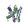
| |||||||||||||||||||||||||||
|---|---|---|---|---|---|---|---|---|---|---|---|---|---|---|---|---|---|---|---|---|---|---|---|---|---|---|---|---|
| 1 |
| |||||||||||||||||||||||||||
| Unit cell |
| |||||||||||||||||||||||||||
| Noncrystallographic symmetry (NCS) | NCS domain:
NCS domain segments:
|
-Components
| #1: Protein | Mass: 35356.797 Da / Num. of mol.: 2 Source method: isolated from a genetically manipulated source Source: (gene. exp.)  Drosophila x virus (isolate Chung/1996) Drosophila x virus (isolate Chung/1996)Strain: isolate Chung/1996 / Production host:  Escherichia coli (E. coli) Escherichia coli (E. coli)References: UniProt: Q96724, Hydrolases; Acting on peptide bonds (peptidases); Serine endopeptidases#2: Chemical | ChemComp-IMD / | Imidazole  Mass: 69.085 Da / Num. of mol.: 1 / Source method: obtained synthetically / Formula: C3H5N2 Mass: 69.085 Da / Num. of mol.: 1 / Source method: obtained synthetically / Formula: C3H5N2#3: Chemical | ChemComp-GOL / | Glycerol  Mass: 92.094 Da / Num. of mol.: 1 / Source method: obtained synthetically / Formula: C3H8O3 Mass: 92.094 Da / Num. of mol.: 1 / Source method: obtained synthetically / Formula: C3H8O3#4: Water | ChemComp-HOH / | Water Mass: 18.015 Da / Num. of mol.: 46 / Source method: isolated from a natural source / Formula: H2O Mass: 18.015 Da / Num. of mol.: 46 / Source method: isolated from a natural source / Formula: H2OHas ligand of interest | N | |
|---|
-Experimental details
-Experiment
| Experiment | Method: X-RAY DIFFRACTION / Number of used crystals: 1 |
|---|
- Sample preparation
| Crystal | Density Matthews: 2.46 Å3/Da / Density % sol: 49.97 % |
|---|---|
| Crystal grow | Temperature: 293 K / Method: vapor diffusion, sitting drop Details: 12% PEG 4K, 200 mM ammonium sulphate and 100 mM sodium acetate pH 4.6 |
-Data collection
| Diffraction | Mean temperature: 100 K / Serial crystal experiment: N |
|---|---|
| Diffraction source | Source: SYNCHROTRON / Site: ESRF  / Beamline: ID29 / Wavelength: 0.97166 Å / Beamline: ID29 / Wavelength: 0.97166 Å |
| Detector | Type: DECTRIS PILATUS 6M / Detector: PIXEL / Date: Jul 3, 2013 |
| Radiation | Protocol: SINGLE WAVELENGTH / Monochromatic (M) / Laue (L): M / Scattering type: x-ray |
| Radiation wavelength | Wavelength: 0.97166 Å / Relative weight: 1 |
| Reflection | Resolution: 1.98→41.6 Å / Num. obs: 8128 / % possible obs: 99.94 % / Redundancy: 7.7 % / CC1/2: 0.999 / Rmerge(I) obs: 0.05459 / Rpim(I) all: 0.02 / Net I/σ(I): 23.01 |
| Reflection shell | Resolution: 1.98→2.051 Å / Num. unique obs: 780 / CC1/2: 0.986 |
- Processing
| Software |
| ||||||||||||||||||||||||||||||||||||||||
|---|---|---|---|---|---|---|---|---|---|---|---|---|---|---|---|---|---|---|---|---|---|---|---|---|---|---|---|---|---|---|---|---|---|---|---|---|---|---|---|---|---|
| Refinement | Method to determine structure: MOLECULAR REPLACEMENT Starting model: 6shw Resolution: 1.98→41.591 Å / SU ML: 0.23 / Cross valid method: THROUGHOUT / σ(F): 1.38 / Phase error: 22.4
| ||||||||||||||||||||||||||||||||||||||||
| Solvent computation | Shrinkage radii: 0.9 Å / VDW probe radii: 1.11 Å | ||||||||||||||||||||||||||||||||||||||||
| Displacement parameters | Biso max: 136.62 Å2 / Biso mean: 46.0206 Å2 / Biso min: 20.97 Å2 | ||||||||||||||||||||||||||||||||||||||||
| Refinement step | Cycle: final / Resolution: 1.98→41.591 Å
| ||||||||||||||||||||||||||||||||||||||||
| Refine LS restraints NCS |
| ||||||||||||||||||||||||||||||||||||||||
| LS refinement shell | Refine-ID: X-RAY DIFFRACTION / Rfactor Rfree error: 0 / % reflection obs: 100 %
| ||||||||||||||||||||||||||||||||||||||||
| Refinement TLS params. | Method: refined / Origin x: -17.6976 Å / Origin y: -4.8716 Å / Origin z: 11.3976 Å
| ||||||||||||||||||||||||||||||||||||||||
| Refinement TLS group |
|
