 Movie
Movie Controller

[English] 日本語
 Yorodumi
- PDB-3nfk: Crystal structure of the PTPN4 PDZ domain complexed with the C-te... -
+ Open data

- Basic information
| Entry | Database: PDB / ID: 3nfk | ||||||
|---|---|---|---|---|---|---|---|
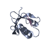
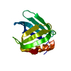
| Title | Crystal structure of the PTPN4 PDZ domain complexed with the C-terminus of a rabies virus G protein | ||||||
 Components Components |
| ||||||
 Keywords Keywords |  PROTEIN BINDING / PDZ-PDZ-binding site complex PROTEIN BINDING / PDZ-PDZ-binding site complex | ||||||
| Function / homology |  Function and homology information Function and homology informationToll Like Receptor 4 (TLR4) Cascade / Interleukin-37 signaling / non-membrane spanning protein tyrosine phosphatase activity / glutamate receptor binding / MECP2 regulates neuronal receptors and channels / cytoskeletal protein binding / protein dephosphorylation / protein-tyrosine-phosphatase / protein tyrosine phosphatase activity / cytoplasmic side of plasma membrane ...Toll Like Receptor 4 (TLR4) Cascade / Interleukin-37 signaling / non-membrane spanning protein tyrosine phosphatase activity / glutamate receptor binding / MECP2 regulates neuronal receptors and channels / cytoskeletal protein binding / protein dephosphorylation / protein-tyrosine-phosphatase / protein tyrosine phosphatase activity / cytoplasmic side of plasma membrane / membrane => GO:0016020 / cytoskeleton / viral envelope / virion membrane / nucleoplasm / cytosol / cytoplasmSimilarity search - Function | ||||||
| Biological species |  Homo sapiens (human) Homo sapiens (human) Rabies virus Rabies virus | ||||||
| Method | X-RAY DIFFRACTION / SYNCHROTRON / MOLECULAR REPLACEMENT / Resolution: 1.43 Å | ||||||
 Authors Authors | Babault, N. / Cordier, F. / Lafage, M. / Cockburn, J. / Haouz, A. / Rey, F.A. / Delepierre, M. / Buc, H. / Lafon, M. / Wolff, N. | ||||||
 Citation Citation | Journal: Structure / Year: 2011 Title: Peptides Targeting the PDZ Domain of PTPN4 Are Efficient Inducers of Glioblastoma Cell Death. Authors: Babault, N. / Cordier, F. / Lafage, M. / Cockburn, J. / Haouz, A. / Prehaud, C. / Rey, F.A. / Delepierre, M. / Buc, H. / Lafon, M. / Wolff, N. | ||||||
| History |
|
- Structure visualization
| Structure viewer | Molecule: MolmilJmol/JSmol |
|---|
- Downloads & links
-Download
| PDBx/mmCIF format | 3nfk.cif.gz | 95.7 KB | Display | PDBx/mmCIF format |
|---|---|---|---|---|
| PDB format | pdb3nfk.ent.gz | 72.5 KB | Display | PDB format |
| PDBx/mmJSON format | 3nfk.json.gz | Tree view | PDBx/mmJSON format | |
| Others |  Other downloads Other downloads |
-Validation report
| Arichive directory | https://data.pdbj.org/pub/pdb/validation_reports/nf/3nfkftp://data.pdbj.org/pub/pdb/validation_reports/nf/3nfk | HTTPS FTP |
|---|
-Related structure data
| Related structure data |  3nflC  2vphS S: Starting model for refinement C: citing same article ( |
|---|---|
| Similar structure data |


-Links
 PDBj
PDBj


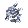
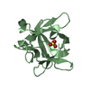
- Assembly
| Deposited unit | 
| ||||||||
|---|---|---|---|---|---|---|---|---|---|
| 1 | 
| ||||||||
| 2 | 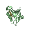
| ||||||||
| 3 | 
| ||||||||
| Unit cell |
|
-Components
| #1: Protein | Mass: 11694.363 Da / Num. of mol.: 2 / Fragment: PDZ domain (UNP residues 499-604) Source method: isolated from a genetically manipulated source Source: (gene. exp.) Homo sapiens (human) / Gene: PTPN4 / Plasmid: pDEST15 / Production host:  Escherichia coli (E. coli) / Strain (production host): BL21 (DE3) star / References: UniProt: P29074 Escherichia coli (E. coli) / Strain (production host): BL21 (DE3) star / References: UniProt: P29074#2: Protein/peptide | Mass: 1476.572 Da / Num. of mol.: 2 / Fragment: UNP residues 512-524 / Source method: obtained synthetically Details: Cyto13-att peptide has been chemically synthetized. The sequence occurs naturally in rabies virus G protein Source: (synth.) Rabies virus / References: UniProt: P03524#3: Chemical | Glycerol  Mass: 92.094 Da / Num. of mol.: 2 / Source method: obtained synthetically / Formula: C3H8O3 Mass: 92.094 Da / Num. of mol.: 2 / Source method: obtained synthetically / Formula: C3H8O3#4: Water | ChemComp-HOH / | Water Mass: 18.015 Da / Num. of mol.: 181 / Source method: isolated from a natural source / Formula: H2O Mass: 18.015 Da / Num. of mol.: 181 / Source method: isolated from a natural source / Formula: H2O |
|---|
-Experimental details
-Experiment
| Experiment | Method: X-RAY DIFFRACTION / Number of used crystals: 1 |
|---|
- Sample preparation
| Crystal | Density Matthews: 2.23 Å3/Da / Density % sol: 44.78 % |
|---|---|
| Crystal grow | Temperature: 291 K / Method: vapor diffusion, hanging drop Details: 24% PEG 1500, 20% Glycerol, VAPOR DIFFUSION, HANGING DROP, temperature 291K |
-Data collection
| Diffraction | Mean temperature: 100 K |
|---|---|
| Diffraction source | Source: SYNCHROTRON / Site: SOLEIL  / Beamline: PROXIMA 1 / Wavelength: 0.918 Å / Beamline: PROXIMA 1 / Wavelength: 0.918 Å |
| Detector | Type: ADSC QUANTUM 315r / Detector: CCD / Date: May 29, 2009 / Details: BI-MORPH MIRRORS |
| Radiation | Monochromator: CRYOGENICALLY COOLED MONOCHROMATOR CRYSTAL / Protocol: SINGLE WAVELENGTH / Monochromatic (M) / Laue (L): M / Scattering type: x-ray |
| Radiation wavelength | Wavelength: 0.918 Å / Relative weight: 1 |
| Reflection | Resolution: 1.43→34.37 Å / Num. all: 44164 / Num. obs: 42795 / % possible obs: 96.9 % / Redundancy: 3.5 % / Biso Wilson estimate: 16.28 Å2 / Rmerge(I) obs: 0.068 / Net I/σ(I): 11 |
| Reflection shell | Resolution: 1.43→1.51 Å / Redundancy: 3.6 % / Rmerge(I) obs: 0.301 / Mean I/σ(I) obs: 4.3 / Num. unique all: 6255 / % possible all: 98.5 |
- Processing
| Software |
| |||||||||||||||||||||||||||||||||||||||||||||||||||||||||||||||||||||||||||||||||||||||||||||||||||||||||||||||||||||||||||||
|---|---|---|---|---|---|---|---|---|---|---|---|---|---|---|---|---|---|---|---|---|---|---|---|---|---|---|---|---|---|---|---|---|---|---|---|---|---|---|---|---|---|---|---|---|---|---|---|---|---|---|---|---|---|---|---|---|---|---|---|---|---|---|---|---|---|---|---|---|---|---|---|---|---|---|---|---|---|---|---|---|---|---|---|---|---|---|---|---|---|---|---|---|---|---|---|---|---|---|---|---|---|---|---|---|---|---|---|---|---|---|---|---|---|---|---|---|---|---|---|---|---|---|---|---|---|---|
| Refinement | Method to determine structure: MOLECULAR REPLACEMENT Starting model: PDB ENTRY 2VPH Resolution: 1.43→14.41 Å / Cor.coef. Fo:Fc: 0.9495 / Cor.coef. Fo:Fc free: 0.9447 / SU R Cruickshank DPI: 0.062 / Cross valid method: THROUGHOUT / σ(F): 0
| |||||||||||||||||||||||||||||||||||||||||||||||||||||||||||||||||||||||||||||||||||||||||||||||||||||||||||||||||||||||||||||
| Displacement parameters | Biso mean: 20.78 Å2
| |||||||||||||||||||||||||||||||||||||||||||||||||||||||||||||||||||||||||||||||||||||||||||||||||||||||||||||||||||||||||||||
| Refine analyze | Luzzati coordinate error obs: 0.175 Å | |||||||||||||||||||||||||||||||||||||||||||||||||||||||||||||||||||||||||||||||||||||||||||||||||||||||||||||||||||||||||||||
| Refinement step | Cycle: LAST / Resolution: 1.43→14.41 Å
| |||||||||||||||||||||||||||||||||||||||||||||||||||||||||||||||||||||||||||||||||||||||||||||||||||||||||||||||||||||||||||||
| Refine LS restraints |
| |||||||||||||||||||||||||||||||||||||||||||||||||||||||||||||||||||||||||||||||||||||||||||||||||||||||||||||||||||||||||||||
| LS refinement shell | Resolution: 1.43→1.47 Å / Total num. of bins used: 20
| |||||||||||||||||||||||||||||||||||||||||||||||||||||||||||||||||||||||||||||||||||||||||||||||||||||||||||||||||||||||||||||
| Refinement TLS params. | Method: refined / Refine-ID: X-RAY DIFFRACTION
| |||||||||||||||||||||||||||||||||||||||||||||||||||||||||||||||||||||||||||||||||||||||||||||||||||||||||||||||||||||||||||||
| Refinement TLS group |
|
