| Entry | Database: PDB / ID: 2vph
|
|---|
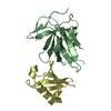
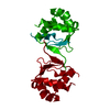
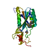
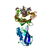
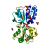
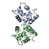
| Title | Crystal structure of the human protein tyrosine phosphatase, non- receptor type 4, PDZ domain |
|---|
 Components Components | TYROSINE-PROTEIN PHOSPHATASE NON-RECEPTOR TYPE 4 |
|---|
 Keywords Keywords |  HYDROLASE / PROTEIN PHOSPHATASE / PTPN4 / PTPMEG / CYTOSKELETON / MEGAKARYOCYTE / DEPHOSPHORYLATION / STRUCTURAL PROTEIN HYDROLASE / PROTEIN PHOSPHATASE / PTPN4 / PTPMEG / CYTOSKELETON / MEGAKARYOCYTE / DEPHOSPHORYLATION / STRUCTURAL PROTEIN |
|---|
| Function / homology |  Function and homology information Function and homology information
Toll Like Receptor 4 (TLR4) Cascade / Interleukin-37 signaling / non-membrane spanning protein tyrosine phosphatase activity / glutamate receptor binding / MECP2 regulates neuronal receptors and channels / cytoskeletal protein binding / protein dephosphorylation / protein-tyrosine-phosphatase / protein tyrosine phosphatase activity / cytoplasmic side of plasma membrane ...Toll Like Receptor 4 (TLR4) Cascade / Interleukin-37 signaling / non-membrane spanning protein tyrosine phosphatase activity / glutamate receptor binding / MECP2 regulates neuronal receptors and channels / cytoskeletal protein binding / protein dephosphorylation / protein-tyrosine-phosphatase / protein tyrosine phosphatase activity / cytoplasmic side of plasma membrane / cytoskeleton / nucleoplasm / cytosol / cytoplasmSimilarity search - Function Protein-tyrosine phosphatase, non-receptor type-3, -4 / PTPN3/4, FERM domain C-lobe / FERM adjacent (FA) / FERM adjacent (FA) / FERM adjacent (FA) / FERM, C-terminal PH-like domain / FERM C-terminal PH-like domain / FERM C-terminal PH-like domain / FERM, N-terminal / FERM N-terminal domain ...Protein-tyrosine phosphatase, non-receptor type-3, -4 / PTPN3/4, FERM domain C-lobe / FERM adjacent (FA) / FERM adjacent (FA) / FERM adjacent (FA) / FERM, C-terminal PH-like domain / FERM C-terminal PH-like domain / FERM C-terminal PH-like domain / FERM, N-terminal / FERM N-terminal domain / FERM domain signature 1. / FERM conserved site / FERM domain signature 2. / FERM central domain / FERM/acyl-CoA-binding protein superfamily / PDZ domain / Pdz3 Domain / FERM central domain / FERM superfamily, second domain / FERM domain / FERM domain profile. / Band 4.1 domain / Band 4.1 homologues / Protein tyrosine phosphatase, catalytic domain / PTP type protein phosphatase domain profile. / Protein-tyrosine phosphatase / Tyrosine-specific protein phosphatase, PTPase domain / Protein-tyrosine phosphatase, catalytic / Protein tyrosine phosphatase, catalytic domain motif / Tyrosine specific protein phosphatases active site. / Protein-tyrosine phosphatase, active site / Tyrosine-specific protein phosphatases domain / Tyrosine specific protein phosphatases domain profile. / Protein-tyrosine phosphatase-like / PDZ domain / PDZ domain profile. / Domain present in PSD-95, Dlg, and ZO-1/2. / PDZ domain / PDZ superfamily / PH-like domain superfamily / Ubiquitin-like domain superfamily / Roll / Mainly BetaSimilarity search - Domain/homology |
|---|
| Biological species |  HOMO SAPIENS (human) HOMO SAPIENS (human) |
|---|
| Method | X-RAY DIFFRACTION / SYNCHROTRON / MOLECULAR REPLACEMENT / Resolution: 1.9 Å |
|---|
 Authors Authors | Roos, A.K. / Wang, J. / Burgess-Brown, N. / Elkins, J.M. / Kavanagh, K. / Pike, A.C.W. / Filippakopoulos, P. / Arrowsmith, C.H. / Weigelt, J. / Edwards, A. ...Roos, A.K. / Wang, J. / Burgess-Brown, N. / Elkins, J.M. / Kavanagh, K. / Pike, A.C.W. / Filippakopoulos, P. / Arrowsmith, C.H. / Weigelt, J. / Edwards, A. / von Delft, F. / Bountra, C. / Knapp, S. |
|---|
 Citation Citation | Journal: To be Published
Title: Crystal Structure of the Human Protein Tyrosine Phosphatase, Non-Receptor Type 4, Pdz Domain
Authors: Roos, A.K. / Wang, J. / Burgess-Brown, N. / Elkins, J.M. / Kavanagh, K. / Pike, A.C.W. / Filippakopoulos, P. / Arrowsmith, C.H. / Weigelt, J. / Edwards, A. / von Delft, F. / Bountra, C. / Knapp, S. |
|---|
| History | | Deposition | Feb 29, 2008 | Deposition site: PDBE / Processing site: PDBE |
|---|
| Revision 1.0 | Mar 18, 2008 | Provider: repository / Type: Initial release |
|---|
| Revision 1.1 | Jul 13, 2011 | Group: Advisory / Version format compliance |
|---|
| Revision 1.2 | Jan 24, 2018 | Group: Database references / Category: citation_author / Item: _citation_author.name |
|---|
| Revision 1.3 | Dec 13, 2023 | Group: Data collection / Database references ...Data collection / Database references / Other / Refinement description
Category: chem_comp_atom / chem_comp_bond ...chem_comp_atom / chem_comp_bond / database_2 / pdbx_database_status / pdbx_initial_refinement_model / struct_ncs_dom_lim
Item: _database_2.pdbx_DOI / _database_2.pdbx_database_accession ..._database_2.pdbx_DOI / _database_2.pdbx_database_accession / _pdbx_database_status.status_code_sf / _struct_ncs_dom_lim.beg_auth_comp_id / _struct_ncs_dom_lim.beg_label_asym_id / _struct_ncs_dom_lim.beg_label_comp_id / _struct_ncs_dom_lim.beg_label_seq_id / _struct_ncs_dom_lim.end_auth_comp_id / _struct_ncs_dom_lim.end_label_asym_id / _struct_ncs_dom_lim.end_label_comp_id / _struct_ncs_dom_lim.end_label_seq_id |
|---|
|
|---|
 Movie
Movie Controller

 Yorodumi
 Open data
 Basic information
 Structure visualization
 Downloads & links
 Other downloads


 PDBj
PDBj


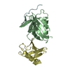
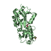
 Assembly
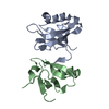


 Mass: 18.015 Da / Num. of mol.: 72 / Source method: isolated from a natural source / Formula: H2O
 Sample preparation
 / Beamline: X10SA / Wavelength: 0.98068
  Processing