 Movie
Movie Controller


+ Open data

- Basic information
| Entry | Database: PDB / ID: 6s36 | ||||||
|---|---|---|---|---|---|---|---|
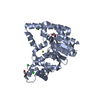
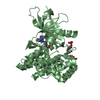
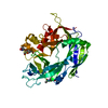
| Title | Crystal structure of E. coli Adenylate kinase R119K mutant | ||||||
 Components Components | Adenylate kinase | ||||||
 Keywords Keywords | TRANSFERASE / ADENYLATE KINASE / R119K VARIANT | ||||||
| Function / homology |  Function and homology information Function and homology informationpurine ribonucleotide interconversion / ADP biosynthetic process / nucleoside monophosphate metabolic process / nucleoside diphosphate metabolic process / adenylate kinase / adenylate kinase activity / AMP salvage / nucleoside diphosphate kinase activity / AMP binding / phosphorylation ...purine ribonucleotide interconversion / ADP biosynthetic process / nucleoside monophosphate metabolic process / nucleoside diphosphate metabolic process / adenylate kinase / adenylate kinase activity / AMP salvage / nucleoside diphosphate kinase activity / AMP binding / phosphorylation / magnesium ion binding / ATP binding / cytosol / cytoplasmSimilarity search - Function | ||||||
| Biological species |  Escherichia coli (E. coli) Escherichia coli (E. coli) | ||||||
| Method | X-RAY DIFFRACTION / SYNCHROTRON / MOLECULAR REPLACEMENT / Resolution: 1.6 Å | ||||||
 Authors Authors | Grundstrom, C. / Rogne, P. / Wolf-Watz, M. / Sauer-Eriksson, A.E. | ||||||
| Funding support |  Sweden, 1items Sweden, 1items
| ||||||
 Citation Citation | Journal: Biochemistry / Year: 2019 Title: Nucleation of an Activating Conformational Change by a Cation-pi Interaction. Authors: Rogne, P. / Andersson, D. / Grundstrom, C. / Sauer-Eriksson, E. / Linusson, A. / Wolf-Watz, M. | ||||||
| History |
|
- Structure visualization
| Structure viewer | Molecule: MolmilJmol/JSmol |
|---|
- Downloads & links
-Download
| PDBx/mmCIF format | 6s36.cif.gz | 106.7 KB | Display | PDBx/mmCIF format |
|---|---|---|---|---|
| PDB format | pdb6s36.ent.gz | 80.6 KB | Display | PDB format |
| PDBx/mmJSON format | 6s36.json.gz | Tree view | PDBx/mmJSON format | |
| Others |  Other downloads Other downloads |
-Validation report
| Arichive directory | https://data.pdbj.org/pub/pdb/validation_reports/s3/6s36ftp://data.pdbj.org/pub/pdb/validation_reports/s3/6s36 | HTTPS FTP |
|---|
-Related structure data
| Related structure data | 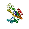 6rzeC 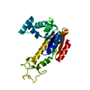 4x8hS S: Starting model for refinement C: citing same article ( |
|---|---|
| Similar structure data |


-Links
 PDBj
PDBj- Assembly
| Deposited unit | 
| ||||||||||||
|---|---|---|---|---|---|---|---|---|---|---|---|---|---|
| 1 |
| ||||||||||||
| Unit cell |
| ||||||||||||
| Components on special symmetry positions |
|
-Components
| #1: Protein | / AK / ATP-AMP transphosphorylase / ATP:AMP phosphotransferase / Adenylate monophosphate kinase Mass: 23592.016 Da / Num. of mol.: 1 Source method: isolated from a genetically manipulated source Source: (gene. exp.) Escherichia coli (E. coli)Gene: adk, D9E35_07195, D9H53_18240, D9H70_06005, D9I87_03740, EB509_06410, EB510_02065, EB515_08900, EC382_09075, ED225_07155, ED607_06260, ED611_06205, ED903_02730, ED944_09135, EEA45_02410, EF173_ ...Gene: adk, D9E35_07195, D9H53_18240, D9H70_06005, D9I87_03740, EB509_06410, EB510_02065, EB515_08900, EC382_09075, ED225_07155, ED607_06260, ED611_06205, ED903_02730, ED944_09135, EEA45_02410, EF173_11005, EIA21_12240, NCTC10444_03756, NCTC9112_04001, NCTC9119_03910, NCTC9969_03944, SAMEA3472056_03545, SAMEA3485101_03900, SAMEA3485113_01288 Production host: Escherichia coli (E. coli)References: UniProt: A0A234NPI7, UniProt: P69441*PLUS, adenylate kinase | ||||||||
|---|---|---|---|---|---|---|---|---|---|
| #2: Chemical | Chloride  Mass: 35.453 Da / Num. of mol.: 3 / Source method: obtained synthetically / Formula: Cl Mass: 35.453 Da / Num. of mol.: 3 / Source method: obtained synthetically / Formula: Cl#3: Chemical | ChemComp-NA / |   Mass: 22.990 Da / Num. of mol.: 1 / Source method: obtained synthetically / Formula: Na Mass: 22.990 Da / Num. of mol.: 1 / Source method: obtained synthetically / Formula: Na#4: Chemical |   Mass: 24.305 Da / Num. of mol.: 2 / Source method: obtained synthetically / Formula: Mg Mass: 24.305 Da / Num. of mol.: 2 / Source method: obtained synthetically / Formula: Mg#5: Water | ChemComp-HOH / | Water Mass: 18.015 Da / Num. of mol.: 240 / Source method: isolated from a natural source / Formula: H2O Mass: 18.015 Da / Num. of mol.: 240 / Source method: isolated from a natural source / Formula: H2OHas ligand of interest | N | |
-Experimental details
-Experiment
| Experiment | Method: X-RAY DIFFRACTION / Number of used crystals: 1 |
|---|
- Sample preparation
| Crystal | Density Matthews: 2.24 Å3/Da / Density % sol: 45.18 % |
|---|---|
| Crystal grow | Temperature: 291 K / Method: vapor diffusion, sitting drop / pH: 8.5 Details: Droplets of 2 to 4 microl protein solution in 30 mM MES pH 6.0 and 50 mM NaCl at 18 mg per ml and 5 molar excess of Ap5A were mixed with 2 microl reservoir solution consisting of 24-30% PEG ...Details: Droplets of 2 to 4 microl protein solution in 30 mM MES pH 6.0 and 50 mM NaCl at 18 mg per ml and 5 molar excess of Ap5A were mixed with 2 microl reservoir solution consisting of 24-30% PEG 4000, 0.2 M MgCl2 and 100 mM Tris-HCl pH 8.5. |
-Data collection
| Diffraction | Mean temperature: 100 K / Serial crystal experiment: N |
|---|---|
| Diffraction source | Source: SYNCHROTRON / Site: ESRF  / Beamline: MASSIF-3 / Wavelength: 0.968 Å / Beamline: MASSIF-3 / Wavelength: 0.968 Å |
| Detector | Type: DECTRIS EIGER X 4M / Detector: PIXEL / Date: Mar 3, 2017 |
| Radiation | Protocol: SINGLE WAVELENGTH / Monochromatic (M) / Laue (L): M / Scattering type: x-ray |
| Radiation wavelength | Wavelength: 0.968 Å / Relative weight: 1 |
| Reflection | Resolution: 1.6→33.3 Å / Num. obs: 27793 / % possible obs: 99.1 % / Redundancy: 6.8 % / CC1/2: 0.998 / Rmerge(I) obs: 0.056 / Rpim(I) all: 0.034 / Net I/σ(I): 16 |
| Reflection shell | Resolution: 1.6→1.63 Å / Rmerge(I) obs: 0.665 / Mean I/σ(I) obs: 2.4 / Num. unique obs: 2806 / CC1/2: 0.775 / Rpim(I) all: 0.411 / % possible all: 99.9 |
- Processing
| Software |
| |||||||||||||||||||||||||||||||||||||||||||||||||||||||||||||||||||||||||||||
|---|---|---|---|---|---|---|---|---|---|---|---|---|---|---|---|---|---|---|---|---|---|---|---|---|---|---|---|---|---|---|---|---|---|---|---|---|---|---|---|---|---|---|---|---|---|---|---|---|---|---|---|---|---|---|---|---|---|---|---|---|---|---|---|---|---|---|---|---|---|---|---|---|---|---|---|---|---|---|
| Refinement | Method to determine structure: MOLECULAR REPLACEMENT Starting model: 4X8H Resolution: 1.6→33.286 Å / SU ML: 0.21 / Cross valid method: FREE R-VALUE / σ(F): 1.35 / Phase error: 25.52
| |||||||||||||||||||||||||||||||||||||||||||||||||||||||||||||||||||||||||||||
| Solvent computation | Shrinkage radii: 0.9 Å / VDW probe radii: 1.11 Å | |||||||||||||||||||||||||||||||||||||||||||||||||||||||||||||||||||||||||||||
| Refinement step | Cycle: LAST / Resolution: 1.6→33.286 Å
| |||||||||||||||||||||||||||||||||||||||||||||||||||||||||||||||||||||||||||||
| Refine LS restraints |
| |||||||||||||||||||||||||||||||||||||||||||||||||||||||||||||||||||||||||||||
| LS refinement shell |
|
