 Movie
Movie Controller


+ Open data

- Basic information
| Entry | Database: PDB / ID: 1ut8 | ||||||
|---|---|---|---|---|---|---|---|
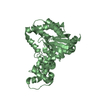
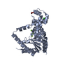
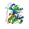
| Title | Divalent metal ions (zinc) bound to T5 5'-exonuclease | ||||||
 Components Components | EXODEOXYRIBONUCLEASE | ||||||
 Keywords Keywords | HYDROLASE / EXONUCLEASE / NUCLEASE | ||||||
| Function / homology |  Function and homology informationviral replication complex / exodeoxyribonuclease (lambda-induced) / late viral transcription / DNA replication, Okazaki fragment processing / double-stranded DNA 5'-3' DNA exonuclease activity / DNA exonuclease activity / double-stranded DNA endonuclease activity / 5'-flap endonuclease activity / 5'-3' exonuclease activity / viral DNA genome replication ...viral replication complex / exodeoxyribonuclease (lambda-induced) / late viral transcription / DNA replication, Okazaki fragment processing / double-stranded DNA 5'-3' DNA exonuclease activity / DNA exonuclease activity / double-stranded DNA endonuclease activity / 5'-flap endonuclease activity / 5'-3' exonuclease activity / viral DNA genome replication / Hydrolases; Acting on ester bonds; Exodeoxyribonucleases producing 5'-phosphomonoesters / 5'-3' DNA exonuclease activity / DNA binding / metal ion binding Function and homology informationviral replication complex / exodeoxyribonuclease (lambda-induced) / late viral transcription / DNA replication, Okazaki fragment processing / double-stranded DNA 5'-3' DNA exonuclease activity / DNA exonuclease activity / double-stranded DNA endonuclease activity / 5'-flap endonuclease activity / 5'-3' exonuclease activity / viral DNA genome replication ...viral replication complex / exodeoxyribonuclease (lambda-induced) / late viral transcription / DNA replication, Okazaki fragment processing / double-stranded DNA 5'-3' DNA exonuclease activity / DNA exonuclease activity / double-stranded DNA endonuclease activity / 5'-flap endonuclease activity / 5'-3' exonuclease activity / viral DNA genome replication / Hydrolases; Acting on ester bonds; Exodeoxyribonucleases producing 5'-phosphomonoesters / 5'-3' DNA exonuclease activity / DNA binding / metal ion bindingSimilarity search - Function | ||||||
| Biological species |  BACTERIOPHAGE T5 (virus) BACTERIOPHAGE T5 (virus) | ||||||
| Method | X-RAY DIFFRACTION / MOLECULAR REPLACEMENT / Resolution: 2.75 Å | ||||||
 Authors Authors | Ceska, T.A. / Sayers, J.R. / Suck, D. | ||||||
 Citation Citation | Journal: Nat.Struct.Mol.Biol. / Year: 2004 Title: Roles of Divalent Metal Ions in Flap Endonuclease-Substrate Interactions Authors: Feng, M. / Patel, D. / Dervan, J. / Ceska, T.A. / Suck, D. / Haq, I. / Sayers, J.R. | ||||||
| History |
|
- Structure visualization
| Structure viewer | Molecule: MolmilJmol/JSmol |
|---|
- Downloads & links
-Download
| PDBx/mmCIF format | 1ut8.cif.gz | 123.8 KB | Display | PDBx/mmCIF format |
|---|---|---|---|---|
| PDB format | pdb1ut8.ent.gz | 96.5 KB | Display | PDB format |
| PDBx/mmJSON format | 1ut8.json.gz | Tree view | PDBx/mmJSON format | |
| Others |  Other downloads Other downloads |
-Validation report
| Arichive directory | https://data.pdbj.org/pub/pdb/validation_reports/ut/1ut8ftp://data.pdbj.org/pub/pdb/validation_reports/ut/1ut8 | HTTPS FTP |
|---|
-Related structure data
| Related structure data | 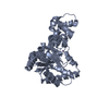 1ut5C 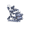 1exnS S: Starting model for refinement C: citing same article ( |
|---|---|
| Similar structure data |


-Links
 PDBj
PDBj

- Assembly
| Deposited unit | 
| ||||||||
|---|---|---|---|---|---|---|---|---|---|
| 1 | 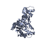
| ||||||||
| 2 | 
| ||||||||
| Unit cell |
|
-Components
| #1: Protein | / 5'-EXONUCLEASE Mass: 33491.867 Da / Num. of mol.: 2 Source method: isolated from a genetically manipulated source Source: (gene. exp.) BACTERIOPHAGE T5 (virus) / Production host:  ESCHERICHIA COLI (E. coli) ESCHERICHIA COLI (E. coli)References: UniProt: P06229, exodeoxyribonuclease (lambda-induced)#2: Chemical |   Mass: 65.409 Da / Num. of mol.: 2 / Source method: obtained synthetically / Formula: Zn Mass: 65.409 Da / Num. of mol.: 2 / Source method: obtained synthetically / Formula: Zn#3: Water | ChemComp-HOH / | Water Mass: 18.015 Da / Num. of mol.: 223 / Source method: isolated from a natural source / Formula: H2O Mass: 18.015 Da / Num. of mol.: 223 / Source method: isolated from a natural source / Formula: H2O |
|---|
-Experimental details
-Experiment
| Experiment | Method: X-RAY DIFFRACTION / Number of used crystals: 1 |
|---|
- Sample preparation
| Crystal | Density Matthews: 3.05 Å3/Da / Density % sol: 65 % | |||||||||||||||||||||||||||||||||||||||||||||||||||||||||||||||
|---|---|---|---|---|---|---|---|---|---|---|---|---|---|---|---|---|---|---|---|---|---|---|---|---|---|---|---|---|---|---|---|---|---|---|---|---|---|---|---|---|---|---|---|---|---|---|---|---|---|---|---|---|---|---|---|---|---|---|---|---|---|---|---|---|
| Crystal grow | pH: 5.6 / Details: 35% AMMONIUM SULFATE, 100MM SODIUM CITRATE PH 5.6 | |||||||||||||||||||||||||||||||||||||||||||||||||||||||||||||||
| Crystal grow | *PLUS pH: 7.5 / Method: vapor diffusion, hanging drop / Details: Ceska, T.A., (1993) J.Mol.Biol., 233, 179. | |||||||||||||||||||||||||||||||||||||||||||||||||||||||||||||||
| Components of the solutions | *PLUS
|
-Data collection
| Diffraction | Mean temperature: 100 K |
|---|---|
| Diffraction source | Source: ROTATING ANODE / Wavelength: 1.5418 |
| Detector | Type: XENTRONICS / Detector: AREA DETECTOR / Date: Nov 15, 1996 |
| Radiation | Monochromator: NI FILTER / Protocol: SINGLE WAVELENGTH / Monochromatic (M) / Laue (L): M / Scattering type: x-ray |
| Radiation wavelength | Wavelength: 1.5418 Å / Relative weight: 1 |
| Reflection | Resolution: 2.75→40 Å / Num. obs: 56234 / % possible obs: 84.5 % / Redundancy: 1.2 % / Rmerge(I) obs: 0.076 |
| Reflection | *PLUS Highest resolution: 2.75 Å / Lowest resolution: 40 Å / Num. obs: 17651 / Num. measured all: 56234 / Rmerge(I) obs: 0.076 |
- Processing
| Software |
| ||||||||||||||||||||||||||||||||||||||||||||||||||||||||||||
|---|---|---|---|---|---|---|---|---|---|---|---|---|---|---|---|---|---|---|---|---|---|---|---|---|---|---|---|---|---|---|---|---|---|---|---|---|---|---|---|---|---|---|---|---|---|---|---|---|---|---|---|---|---|---|---|---|---|---|---|---|---|
| Refinement | Method to determine structure: MOLECULAR REPLACEMENT Starting model: PDB ENTRY 1EXN Resolution: 2.75→25 Å / Cross valid method: THROUGHOUT / σ(F): 0
| ||||||||||||||||||||||||||||||||||||||||||||||||||||||||||||
| Solvent computation | Solvent model: MASK / Bsol: 66.2 Å2 / ksol: 0.4 e/Å3 | ||||||||||||||||||||||||||||||||||||||||||||||||||||||||||||
| Refinement step | Cycle: LAST / Resolution: 2.75→25 Å
| ||||||||||||||||||||||||||||||||||||||||||||||||||||||||||||
| Refine LS restraints |
| ||||||||||||||||||||||||||||||||||||||||||||||||||||||||||||
| Xplor file |
| ||||||||||||||||||||||||||||||||||||||||||||||||||||||||||||
| Refinement | *PLUS Lowest resolution: 40 Å / % reflection Rfree: 10 % | ||||||||||||||||||||||||||||||||||||||||||||||||||||||||||||
| Solvent computation | *PLUS | ||||||||||||||||||||||||||||||||||||||||||||||||||||||||||||
| Displacement parameters | *PLUS |
