 Movie
Movie Controller


+ Open data

- Basic information
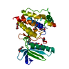
| Entry | Database: PDB / ID: 1xo1 | ||||||
|---|---|---|---|---|---|---|---|
| Title | T5 5'-EXONUCLEASE MUTANT K83A | ||||||
 Components Components | 5'-EXONUCLEASE | ||||||
 Keywords Keywords |  HYDROLASE / EXONUCLEASE / NUCLEASE HYDROLASE / EXONUCLEASE / NUCLEASE | ||||||
| Function / homology |  Function and homology informationviral replication complex / exodeoxyribonuclease (lambda-induced) / late viral transcription / DNA replication, Okazaki fragment processing / double-stranded DNA 5'-3' DNA exonuclease activity / DNA exonuclease activity / double-stranded DNA endonuclease activity / 5'-flap endonuclease activity / 5'-3' exonuclease activity / viral DNA genome replication ...viral replication complex / exodeoxyribonuclease (lambda-induced) / late viral transcription / DNA replication, Okazaki fragment processing / double-stranded DNA 5'-3' DNA exonuclease activity / DNA exonuclease activity / double-stranded DNA endonuclease activity / 5'-flap endonuclease activity / 5'-3' exonuclease activity / viral DNA genome replication / Hydrolases; Acting on ester bonds; Exodeoxyribonucleases producing 5'-phosphomonoesters / 5'-3' DNA exonuclease activity / DNA binding / metal ion binding Function and homology informationviral replication complex / exodeoxyribonuclease (lambda-induced) / late viral transcription / DNA replication, Okazaki fragment processing / double-stranded DNA 5'-3' DNA exonuclease activity / DNA exonuclease activity / double-stranded DNA endonuclease activity / 5'-flap endonuclease activity / 5'-3' exonuclease activity / viral DNA genome replication ...viral replication complex / exodeoxyribonuclease (lambda-induced) / late viral transcription / DNA replication, Okazaki fragment processing / double-stranded DNA 5'-3' DNA exonuclease activity / DNA exonuclease activity / double-stranded DNA endonuclease activity / 5'-flap endonuclease activity / 5'-3' exonuclease activity / viral DNA genome replication / Hydrolases; Acting on ester bonds; Exodeoxyribonucleases producing 5'-phosphomonoesters / 5'-3' DNA exonuclease activity / DNA binding / metal ion bindingSimilarity search - Function | ||||||
| Biological species |  Enterobacteria phage T5 (virus) Enterobacteria phage T5 (virus) | ||||||
| Method | X-RAY DIFFRACTION / MOLECULAR REPLACEMENT / Resolution: 2.5 Å | ||||||
 Authors Authors | Ceska, T.A. / Suck, D. / Sayers, J.R. | ||||||
 Citation Citation | Journal: Proc.Natl.Acad.Sci.USA / Year: 1999 Title: Mutagenesis of conserved lysine residues in bacteriophage T5 5'-3' exonuclease suggests separate mechanisms of endo-and exonucleolytic cleavage. Authors: Garforth, S.J. / Ceska, T.A. / Suck, D. / Sayers, J.R. #1: Journal: Trends Biochem.Sci. / Year: 1998Title: Structure-Specific DNA Cleavage by 5' Nucleases Authors: Ceska, T.A. / Sayers, J.R. #2: Journal: Nature / Year: 1996Title: A Helical Arch Allowing Single-Stranded DNA to Thread Through T5 5'-Exonuclease Authors: Ceska, T.A. / Sayers, J.R. / Stier, G. / Suck, D. #3: Journal: J.Biol.Chem. / Year: 1990Title: Properties of Overexpressed Phage T5 D15 Exonuclease. Similarities with Escherichia Coli DNA Polymerase I 5'-3' Exonuclease Authors: Sayers, J.R. / Eckstein, F. | ||||||
| History |
|
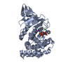
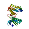
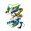
- Structure visualization
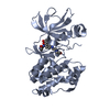
| Structure viewer | Molecule: MolmilJmol/JSmol |
|---|
- Downloads & links
-Download
| PDBx/mmCIF format | 1xo1.cif.gz | 115.3 KB | Display | PDBx/mmCIF format |
|---|---|---|---|---|
| PDB format | pdb1xo1.ent.gz | 88.7 KB | Display | PDB format |
| PDBx/mmJSON format | 1xo1.json.gz | Tree view | PDBx/mmJSON format | |
| Others |  Other downloads Other downloads |
-Validation report
| Arichive directory | https://data.pdbj.org/pub/pdb/validation_reports/xo/1xo1ftp://data.pdbj.org/pub/pdb/validation_reports/xo/1xo1 | HTTPS FTP |
|---|
-Related structure data
| Related structure data | 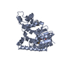 1exnS S: Starting model for refinement |
|---|---|
| Similar structure data |


-Links
 PDBj
PDBj

- Assembly
| Deposited unit | 
| ||||||||||
|---|---|---|---|---|---|---|---|---|---|---|---|
| 1 | 
| ||||||||||
| 2 | 
| ||||||||||
| Unit cell |
| ||||||||||
| Noncrystallographic symmetry (NCS) | NCS oper: (Code: given Matrix: (-1, 0.0025, 0.0074), Vector : |
-Components
| #1: Protein | Mass: 33433.766 Da / Num. of mol.: 2 / Mutation: K83A Source method: isolated from a genetically manipulated source Source: (gene. exp.) Enterobacteria phage T5 (virus) / Genus: T5-like viruses / Strain: M72 / Gene: D15 / Gene (production host): D15 / Production host:  Escherichia coli (E. coli) / Strain (production host): M72 Escherichia coli (E. coli) / Strain (production host): M72References: UniProt: P06229, exodeoxyribonuclease (lambda-induced)#2: Water | ChemComp-HOH / | Water Mass: 18.015 Da / Num. of mol.: 316 / Source method: isolated from a natural source / Formula: H2O Mass: 18.015 Da / Num. of mol.: 316 / Source method: isolated from a natural source / Formula: H2O |
|---|
-Experimental details
-Experiment
| Experiment | Method: X-RAY DIFFRACTION / Number of used crystals: 1 |
|---|
- Sample preparation
| Crystal | Density Matthews: 2.31 Å3/Da / Density % sol: 46.67 % | |||||||||||||||||||||||||||||||||||||||||||||||||||||||||||||||
|---|---|---|---|---|---|---|---|---|---|---|---|---|---|---|---|---|---|---|---|---|---|---|---|---|---|---|---|---|---|---|---|---|---|---|---|---|---|---|---|---|---|---|---|---|---|---|---|---|---|---|---|---|---|---|---|---|---|---|---|---|---|---|---|---|
| Crystal grow | pH: 5.4 / Details: pH 5.4 | |||||||||||||||||||||||||||||||||||||||||||||||||||||||||||||||
| Crystal grow | *PLUS Method: vapor diffusion, hanging drop / Details: Ceska, T.A., (1993) J.Mol.Biol., 233, 179. / pH: 7.5 | |||||||||||||||||||||||||||||||||||||||||||||||||||||||||||||||
| Components of the solutions | *PLUS
|
-Data collection
| Diffraction | Mean temperature: 100 K |
|---|---|
| Diffraction source | Wavelength: 1.5418 |
| Detector | Type: XENTRONICS / Detector: AREA DETECTOR / Details: MIRRORS |
| Radiation | Protocol: SINGLE WAVELENGTH / Monochromatic (M) / Laue (L): M / Scattering type: x-ray |
| Radiation wavelength | Wavelength: 1.5418 Å / Relative weight: 1 |
| Reflection | Resolution: 2.5→25 Å / Num. all: 21915 / Num. obs: 21915 / % possible obs: 99.6 % / Observed criterion σ(I): 0 / Redundancy: 7.2 % / Rsym value: 8.2 |
| Reflection | *PLUS Num. measured all: 158564 / Rmerge(I) obs: 0.082 |
- Processing
| Software |
| ||||||||||||||||||||||||||||||||||||||||||||||||||||||||||||
|---|---|---|---|---|---|---|---|---|---|---|---|---|---|---|---|---|---|---|---|---|---|---|---|---|---|---|---|---|---|---|---|---|---|---|---|---|---|---|---|---|---|---|---|---|---|---|---|---|---|---|---|---|---|---|---|---|---|---|---|---|---|
| Refinement | Method to determine structure: MOLECULAR REPLACEMENT Starting model: 1EXN Resolution: 2.5→6 Å / Data cutoff high absF: 10000000 / Data cutoff low absF: 0.001 / Isotropic thermal model: RESTRAINED / Cross valid method: THROUGHOUT / σ(F): 2
| ||||||||||||||||||||||||||||||||||||||||||||||||||||||||||||
| Displacement parameters | Biso mean: 25 Å2 | ||||||||||||||||||||||||||||||||||||||||||||||||||||||||||||
| Refinement step | Cycle: LAST / Resolution: 2.5→6 Å
| ||||||||||||||||||||||||||||||||||||||||||||||||||||||||||||
| Refine LS restraints |
| ||||||||||||||||||||||||||||||||||||||||||||||||||||||||||||
| Refine LS restraints NCS | NCS model details: RESTRAINTS | ||||||||||||||||||||||||||||||||||||||||||||||||||||||||||||
| Xplor file | Serial no: 1 / Param file: PARHCSDX.PRO / Topol file: TOPHCSDX.PRO | ||||||||||||||||||||||||||||||||||||||||||||||||||||||||||||
| Software | *PLUS Name: X-PLOR / Version: 3.1 / Classification: refinement | ||||||||||||||||||||||||||||||||||||||||||||||||||||||||||||
| Refinement | *PLUS Highest resolution: 2.5 Å / Lowest resolution: 6 Å / σ(F): 2 / % reflection Rfree: 10 % / Rfactor obs: 0.226 | ||||||||||||||||||||||||||||||||||||||||||||||||||||||||||||
| Solvent computation | *PLUS | ||||||||||||||||||||||||||||||||||||||||||||||||||||||||||||
| Displacement parameters | *PLUS Biso mean: 25 Å2 | ||||||||||||||||||||||||||||||||||||||||||||||||||||||||||||
| Refine LS restraints | *PLUS
|
