 Movie
Movie Controller

[English] 日本語
 Yorodumi
- PDB-6ruq: Structure of GluA2cryst in complex the antagonist ZK200775 and th... -
+ Open data

- Basic information
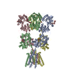
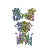
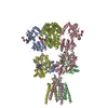
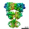
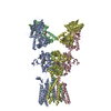
| Entry | Database: PDB / ID: 6ruq | ||||||||||||||||||
|---|---|---|---|---|---|---|---|---|---|---|---|---|---|---|---|---|---|---|---|
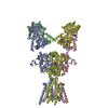
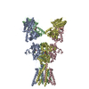
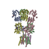
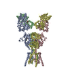
| Title | Structure of GluA2cryst in complex the antagonist ZK200775 and the negative allosteric modulator GYKI53655 at 4.65 A resolution | ||||||||||||||||||
 Components Components | Glutamate receptor 2 GRIA2 GRIA2 | ||||||||||||||||||
 Keywords Keywords | MEMBRANE PROTEIN / Receptor Negative allosteric modulator Antagonist Non-competitve antagonist | ||||||||||||||||||
| Function / homology |  Function and homology information Function and homology informationspine synapse / dendritic spine neck / dendritic spine head / Activation of AMPA receptors / response to lithium ion / perisynaptic space / cellular response to glycine / AMPA glutamate receptor activity / Trafficking of GluR2-containing AMPA receptors / immunoglobulin binding ...spine synapse / dendritic spine neck / dendritic spine head / Activation of AMPA receptors / response to lithium ion / perisynaptic space / cellular response to glycine / AMPA glutamate receptor activity / Trafficking of GluR2-containing AMPA receptors / immunoglobulin binding / AMPA glutamate receptor complex / kainate selective glutamate receptor activity / ionotropic glutamate receptor complex / extracellularly glutamate-gated ion channel activity / asymmetric synapse / regulation of receptor recycling / Unblocking of NMDA receptors, glutamate binding and activation / glutamate receptor binding / positive regulation of synaptic transmission / glutamate-gated receptor activity / presynaptic active zone membrane / response to fungicide / regulation of synaptic transmission, glutamatergic / cellular response to brain-derived neurotrophic factor stimulus / somatodendritic compartment / dendrite membrane / ligand-gated monoatomic ion channel activity involved in regulation of presynaptic membrane potential / ionotropic glutamate receptor binding / ionotropic glutamate receptor signaling pathway / dendrite cytoplasm / cytoskeletal protein binding / SNARE binding / dendritic shaft / transmitter-gated monoatomic ion channel activity involved in regulation of postsynaptic membrane potential / synaptic membrane / synaptic transmission, glutamatergic / PDZ domain binding / postsynaptic density membrane / protein tetramerization / modulation of chemical synaptic transmission / Schaffer collateral - CA1 synapse / establishment of protein localization / terminal bouton / receptor internalization / synaptic vesicle membrane / cerebral cortex development / synaptic vesicle / presynapse / signaling receptor activity / presynaptic membrane / amyloid-beta binding / growth cone / perikaryon / chemical synaptic transmission / postsynaptic membrane / scaffold protein binding / dendritic spine / postsynaptic density / neuron projection / axon / dendrite / neuronal cell body / glutamatergic synapse / synapse / protein-containing complex binding / endoplasmic reticulum membrane / protein kinase binding / cell surface / endoplasmic reticulum / protein-containing complex / membrane / identical protein binding / plasma membraneSimilarity search - Function | ||||||||||||||||||
| Biological species |  Rattus norvegicus (Norway rat) Rattus norvegicus (Norway rat) | ||||||||||||||||||
| Method | X-RAY DIFFRACTION / SYNCHROTRON / MOLECULAR REPLACEMENT / molecular replacement / Resolution: 4.65 Å | ||||||||||||||||||
 Authors Authors | Krintel, C. / Venskutonyte, R. / Mirza, O.A. / Gajhede, M. / Kastrup, J.S. | ||||||||||||||||||
| Funding support |  Denmark, 5items Denmark, 5items
| ||||||||||||||||||
 Citation Citation | Journal: FEBS J / Year: 2021 Title: Binding of a negative allosteric modulator and competitive antagonist can occur simultaneously at the ionotropic glutamate receptor GluA2. Authors: Christian Krintel / Jerzy Dorosz / Andreas Haahr Larsen / Thor Seneca Thorsen / Raminta Venskutonytė / Osman Mirza / Michael Gajhede / Thomas Boesen / Jette Sandholm Kastrup /  Abstract: Ionotropic glutamate receptors are ligand-gated ion channels governing neurotransmission in the central nervous system. Three major types of antagonists are known for the AMPA-type receptor GluA2: ...Ionotropic glutamate receptors are ligand-gated ion channels governing neurotransmission in the central nervous system. Three major types of antagonists are known for the AMPA-type receptor GluA2: competitive, noncompetitive (i.e., negative allosteric modulators; NAMs) used for treatment of epilepsy, and uncompetitive antagonists. We here report a 4.65 Å resolution X-ray structure of GluA2, revealing that four molecules of the competitive antagonist ZK200775 and four molecules of the NAM GYKI53655 are capable of binding at the same time. Using negative stain electron microscopy, we show that GYKI53655 alone or ZK200775/GYKI53655 in combination predominantly results in compact receptor forms. The agonist AMPA provides a mixed population of compact and bulgy shapes of GluA2 not impacted by addition of GYKI53655. Taken together, this suggests that the two different mechanisms of antagonism that lead to channel closure are independent and that the distribution between bulgy and compact receptors primarily depends on the ligand bound in the glutamate binding site. DATABASE: The atomic coordinates and structure factors from the crystal structure determination have been deposited in the Protein Data Bank under accession code https://doi.org/10.2210/pdb6RUQ/pdb. The electron microscopy 3D reconstruction volumes have been deposited in EMDB (EMD-4875: Apo; EMD-4920: ZK200775/GYKI53655; EMD-4921: AMPA compact; EMD-4922: AMPA/GYKI53655 bulgy; EMD-4923: GYKI53655; EMD-4924: AMPA bulgy; EMD-4925: AMPA/GYKI53655 compact). | ||||||||||||||||||
| History |
|
- Structure visualization
| Structure viewer | Molecule: MolmilJmol/JSmol |
|---|
- Downloads & links
-Download
| PDBx/mmCIF format | 6ruq.cif.gz | 768.9 KB | Display | PDBx/mmCIF format |
|---|---|---|---|---|
| PDB format | pdb6ruq.ent.gz | 512.6 KB | Display | PDB format |
| PDBx/mmJSON format | 6ruq.json.gz | Tree view | PDBx/mmJSON format | |
| Others |  Other downloads Other downloads |
-Validation report
| Arichive directory | https://data.pdbj.org/pub/pdb/validation_reports/ru/6ruqftp://data.pdbj.org/pub/pdb/validation_reports/ru/6ruq | HTTPS FTP |
|---|
-Related structure data
| Related structure data |  4875C 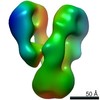 4920C  4921C  4922C 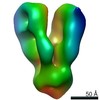 4923C  4924C 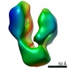 4925C  5l1hS S: Starting model for refinement C: citing same article ( |
|---|---|
| Similar structure data |
-Links
 PDBj
PDBj


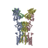
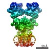
- Assembly
| Deposited unit | 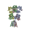
| ||||||||||||||||||||||||||||||||||||||||||||||||||||||||||||||||||||||||||||||||||||||||||||||
|---|---|---|---|---|---|---|---|---|---|---|---|---|---|---|---|---|---|---|---|---|---|---|---|---|---|---|---|---|---|---|---|---|---|---|---|---|---|---|---|---|---|---|---|---|---|---|---|---|---|---|---|---|---|---|---|---|---|---|---|---|---|---|---|---|---|---|---|---|---|---|---|---|---|---|---|---|---|---|---|---|---|---|---|---|---|---|---|---|---|---|---|---|---|---|---|
| 1 |
| ||||||||||||||||||||||||||||||||||||||||||||||||||||||||||||||||||||||||||||||||||||||||||||||
| Unit cell |
| ||||||||||||||||||||||||||||||||||||||||||||||||||||||||||||||||||||||||||||||||||||||||||||||
| Noncrystallographic symmetry (NCS) | NCS domain:
NCS domain segments: Ens-ID: 1
|