 Movie
Movie Controller

[English] 日本語
 Yorodumi
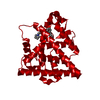
- PDB-6rup: Human mitochondrial single-stranded DNA binding protein, SSBP1, a... -
+ Open data

- Basic information
| Entry | Database: PDB / ID: 6rup | ||||||
|---|---|---|---|---|---|---|---|
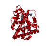
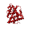
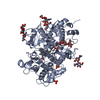
| Title | Human mitochondrial single-stranded DNA binding protein, SSBP1, at 2.1 A resolution - elucidated sequence | ||||||
 Components Components |
| ||||||
 Keywords Keywords |  DNA BINDING PROTEIN / Single-stranded DNA binding protein / mitochondria / mtDNA / mtDNA replication / mitochondrial RNA granules DNA BINDING PROTEIN / Single-stranded DNA binding protein / mitochondria / mtDNA / mtDNA replication / mitochondrial RNA granules | ||||||
| Function / homology |  Function and homology information Function and homology informationpositive regulation of mitochondrial DNA replication / positive regulation of helicase activity / mitochondrial DNA replication / DNA unwinding involved in DNA replication / mitochondrial nucleoid / Transcriptional activation of mitochondrial biogenesis / single-stranded DNA binding / protein homotetramerization / mitochondrial matrix / chromatin binding ...positive regulation of mitochondrial DNA replication / positive regulation of helicase activity / mitochondrial DNA replication / DNA unwinding involved in DNA replication / mitochondrial nucleoid / Transcriptional activation of mitochondrial biogenesis / single-stranded DNA binding / protein homotetramerization / mitochondrial matrix / chromatin binding / protein homodimerization activity / mitochondrion / RNA binding / extracellular exosome / identical protein binding / nucleusSimilarity search - Function | ||||||
| Biological species |  Homo sapiens (human) Homo sapiens (human) | ||||||
| Method | X-RAY DIFFRACTION / SYNCHROTRON / MOLECULAR REPLACEMENT / Resolution: 2.1 Å | ||||||
 Authors Authors | Tarres-Sole, A. / Chakraborty, A. / Spelbrink, H.N. / Delettre, C. / Sola, M. | ||||||
| Funding support |  Spain, 1items Spain, 1items
| ||||||
 Citation Citation | Journal: J.Clin.Invest. / Year: 2020 Title: Dominant mutations in mtDNA maintenance gene SSBP1 cause optic atrophy and foveopathy. Authors: Piro-Megy, C. / Sarzi, E. / Tarres-Sole, A. / Pequignot, M. / Hensen, F. / Quiles, M. / Manes, G. / Chakraborty, A. / Senechal, A. / Bocquet, B. / Cazevieille, C. / Roubertie, A. / Muller, A. ...Authors: Piro-Megy, C. / Sarzi, E. / Tarres-Sole, A. / Pequignot, M. / Hensen, F. / Quiles, M. / Manes, G. / Chakraborty, A. / Senechal, A. / Bocquet, B. / Cazevieille, C. / Roubertie, A. / Muller, A. / Charif, M. / Goudenege, D. / Lenaers, G. / Wilhelm, H. / Kellner, U. / Weisschuh, N. / Wissinger, B. / Zanlonghi, X. / Hamel, C. / Spelbrink, J.N. / Sola, M. / Delettre, C. #1: Journal: Nucleic Acids Res. / Year: 2019 Title: Mitochondrial RNA granules are critically dependent on mtDNA replication factors Twinkle and mtSSB. Authors: Hensen, F. / Potter, A. / van Esveld, S.L. / Tarres-Sole, A. / Chakraborty, A. / Sola, M. / Spelbrink, J.N. | ||||||
| History |
|
- Structure visualization
| Structure viewer | Molecule: MolmilJmol/JSmol |
|---|
- Downloads & links
-Download
| PDBx/mmCIF format | 6rup.cif.gz | 107.8 KB | Display | PDBx/mmCIF format |
|---|---|---|---|---|
| PDB format | pdb6rup.ent.gz | 82.8 KB | Display | PDB format |
| PDBx/mmJSON format | 6rup.json.gz | Tree view | PDBx/mmJSON format | |
| Others |  Other downloads Other downloads |
-Validation report
| Arichive directory | https://data.pdbj.org/pub/pdb/validation_reports/ru/6rupftp://data.pdbj.org/pub/pdb/validation_reports/ru/6rup | HTTPS FTP |
|---|
-Related structure data
| Related structure data | 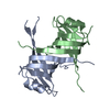 3ullS S: Starting model for refinement |
|---|---|
| Similar structure data |


-Links
 PDBj
PDBj- Assembly
| Deposited unit | 
| ||||||||
|---|---|---|---|---|---|---|---|---|---|
| 1 | 
| ||||||||
| Unit cell |
| ||||||||
| Components on special symmetry positions |
|
-Components
| #1: Protein | Mass: 16489.557 Da / Num. of mol.: 2 Source method: isolated from a genetically manipulated source Source: (gene. exp.) Homo sapiens (human) / Gene: SSBP1, SSBP / Plasmid: pCRI7a / Production host:  Escherichia coli (E. coli) / Variant (production host): codon+ / References: UniProt: Q04837 Escherichia coli (E. coli) / Variant (production host): codon+ / References: UniProt: Q04837#2: Protein/peptide | | Mass: 366.326 Da / Num. of mol.: 1 Source method: isolated from a genetically manipulated source Source: (gene. exp.) Homo sapiens (human) / Production host: Escherichia coli (E. coli)#3: Chemical |   Mass: 24.305 Da / Num. of mol.: 2 / Source method: obtained synthetically / Formula: Mg Mass: 24.305 Da / Num. of mol.: 2 / Source method: obtained synthetically / Formula: Mg#4: Water | ChemComp-HOH / | Water Mass: 18.015 Da / Num. of mol.: 95 / Source method: isolated from a natural source / Formula: H2O Mass: 18.015 Da / Num. of mol.: 95 / Source method: isolated from a natural source / Formula: H2O |
|---|
-Experimental details
-Experiment
| Experiment | Method: X-RAY DIFFRACTION / Number of used crystals: 1 |
|---|
- Sample preparation
| Crystal grow | Temperature: 293 K / Method: evaporation / pH: 6.5 Details: 11 % PEG1500, 0.1 M cacodylate pH 6.5, 0.2 M magnesium chloride. cryoprotectant solution included |
|---|
-Data collection
| Diffraction | Mean temperature: 100 K / Serial crystal experiment: N |
|---|---|
| Diffraction source | Source: SYNCHROTRON / Site: ALBA / Beamline: XALOC / Wavelength: 0.9795 Å |
| Detector | Type: DECTRIS PILATUS3 R CdTe 300K / Detector: PIXEL / Date: Oct 28, 2016 |
| Radiation | Protocol: SINGLE WAVELENGTH / Monochromatic (M) / Laue (L): M / Scattering type: x-ray |
| Radiation wavelength | Wavelength: 0.9795 Å / Relative weight: 1 |
| Reflection | Resolution: 2.1→45.58 Å / Num. obs: 14943 / % possible obs: 99.5 % / Observed criterion σ(I): 1.09 / Redundancy: 11 % / Biso Wilson estimate: 52.63 Å2 / CC1/2: 0.999 / Rrim(I) all: 0.096 / Net I/σ(I): 16.28 |
| Reflection shell | Resolution: 2.1→2.23 Å / Redundancy: 11.2 % / Num. unique obs: 2342 / CC1/2: 0.542 / Rrim(I) all: 0.245 / % possible all: 97.5 |
- Processing
| Software |
| ||||||||||||||||||||||||||||||||||||||||||||||||||||||||||||||||||||||||||||||||||||||||||||||||||||||||||||||||||||||||||||||||||||||||||||||||||||||||||||||||||||||||||||||||||||||||||||||||||||||||||||||||||||||||||||||||||||||||||||||||||||||||||||||||||||||||||||||||||||||||||||||||||||||||||||
|---|---|---|---|---|---|---|---|---|---|---|---|---|---|---|---|---|---|---|---|---|---|---|---|---|---|---|---|---|---|---|---|---|---|---|---|---|---|---|---|---|---|---|---|---|---|---|---|---|---|---|---|---|---|---|---|---|---|---|---|---|---|---|---|---|---|---|---|---|---|---|---|---|---|---|---|---|---|---|---|---|---|---|---|---|---|---|---|---|---|---|---|---|---|---|---|---|---|---|---|---|---|---|---|---|---|---|---|---|---|---|---|---|---|---|---|---|---|---|---|---|---|---|---|---|---|---|---|---|---|---|---|---|---|---|---|---|---|---|---|---|---|---|---|---|---|---|---|---|---|---|---|---|---|---|---|---|---|---|---|---|---|---|---|---|---|---|---|---|---|---|---|---|---|---|---|---|---|---|---|---|---|---|---|---|---|---|---|---|---|---|---|---|---|---|---|---|---|---|---|---|---|---|---|---|---|---|---|---|---|---|---|---|---|---|---|---|---|---|---|---|---|---|---|---|---|---|---|---|---|---|---|---|---|---|---|---|---|---|---|---|---|---|---|---|---|---|---|---|---|---|---|---|---|---|---|---|---|---|---|---|---|---|---|---|---|---|---|---|---|---|---|---|---|---|---|---|---|---|---|---|---|---|---|---|---|---|---|---|---|---|---|---|---|---|---|---|---|---|---|---|---|
| Refinement | Method to determine structure: MOLECULAR REPLACEMENT Starting model: 3ULL Resolution: 2.1→45.58 Å / Cor.coef. Fo:Fc: 0.949 / Cor.coef. Fo:Fc free: 0.935 / SU R Cruickshank DPI: 0.21 / Cross valid method: THROUGHOUT / σ(F): 0 / SU R Blow DPI: 0.228 / SU Rfree Blow DPI: 0.19 / SU Rfree Cruickshank DPI: 0.185
| ||||||||||||||||||||||||||||||||||||||||||||||||||||||||||||||||||||||||||||||||||||||||||||||||||||||||||||||||||||||||||||||||||||||||||||||||||||||||||||||||||||||||||||||||||||||||||||||||||||||||||||||||||||||||||||||||||||||||||||||||||||||||||||||||||||||||||||||||||||||||||||||||||||||||||||
| Displacement parameters |
| ||||||||||||||||||||||||||||||||||||||||||||||||||||||||||||||||||||||||||||||||||||||||||||||||||||||||||||||||||||||||||||||||||||||||||||||||||||||||||||||||||||||||||||||||||||||||||||||||||||||||||||||||||||||||||||||||||||||||||||||||||||||||||||||||||||||||||||||||||||||||||||||||||||||||||||
| Refine analyze | Luzzati coordinate error obs: 0.28 Å | ||||||||||||||||||||||||||||||||||||||||||||||||||||||||||||||||||||||||||||||||||||||||||||||||||||||||||||||||||||||||||||||||||||||||||||||||||||||||||||||||||||||||||||||||||||||||||||||||||||||||||||||||||||||||||||||||||||||||||||||||||||||||||||||||||||||||||||||||||||||||||||||||||||||||||||
| Refinement step | Cycle: 1 / Resolution: 2.1→45.58 Å
| ||||||||||||||||||||||||||||||||||||||||||||||||||||||||||||||||||||||||||||||||||||||||||||||||||||||||||||||||||||||||||||||||||||||||||||||||||||||||||||||||||||||||||||||||||||||||||||||||||||||||||||||||||||||||||||||||||||||||||||||||||||||||||||||||||||||||||||||||||||||||||||||||||||||||||||
| Refine LS restraints |
| ||||||||||||||||||||||||||||||||||||||||||||||||||||||||||||||||||||||||||||||||||||||||||||||||||||||||||||||||||||||||||||||||||||||||||||||||||||||||||||||||||||||||||||||||||||||||||||||||||||||||||||||||||||||||||||||||||||||||||||||||||||||||||||||||||||||||||||||||||||||||||||||||||||||||||||
| LS refinement shell | Resolution: 2.1→2.27 Å / Rfactor Rfree error: 0 / Total num. of bins used: 7
| ||||||||||||||||||||||||||||||||||||||||||||||||||||||||||||||||||||||||||||||||||||||||||||||||||||||||||||||||||||||||||||||||||||||||||||||||||||||||||||||||||||||||||||||||||||||||||||||||||||||||||||||||||||||||||||||||||||||||||||||||||||||||||||||||||||||||||||||||||||||||||||||||||||||||||||
| Refinement TLS params. | Method: refined / Refine-ID: X-RAY DIFFRACTION
| ||||||||||||||||||||||||||||||||||||||||||||||||||||||||||||||||||||||||||||||||||||||||||||||||||||||||||||||||||||||||||||||||||||||||||||||||||||||||||||||||||||||||||||||||||||||||||||||||||||||||||||||||||||||||||||||||||||||||||||||||||||||||||||||||||||||||||||||||||||||||||||||||||||||||||||
| Refinement TLS group |
|
