 Movie
Movie Controller

[English] 日本語
 Yorodumi
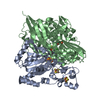
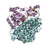
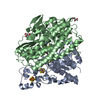
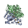
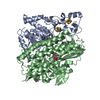
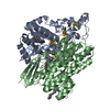
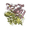
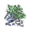
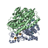
- PDB-6rtp: THE 3D STRUCTURE OF [NIFESE] HYDROGENASE G50T VaRIANT FROM DESULF... -
+ Open data

- Basic information
| Entry | Database: PDB / ID: 6rtp | |||||||||
|---|---|---|---|---|---|---|---|---|---|---|
| Title | THE 3D STRUCTURE OF [NIFESE] HYDROGENASE G50T VaRIANT FROM DESULFOVIBRIO VULGARIS HILDENBOROUGH AT 1.10 ANGSTROM RESOLUTION | |||||||||
 Components Components | (Periplasmic [NiFeSe] hydrogenase, ...) x 2 | |||||||||
 Keywords Keywords |  OXIDOREDUCTASE / NIFESE-SITE / H2 CLEAVAGE/PRODUCTION / O2 TOLERANCE OXIDOREDUCTASE / NIFESE-SITE / H2 CLEAVAGE/PRODUCTION / O2 TOLERANCE | |||||||||
| Function / homology |  Function and homology informationcytochrome-c3 hydrogenase / ferredoxin hydrogenase / cytochrome-c3 hydrogenase activity / ferredoxin hydrogenase complex / ferredoxin hydrogenase activity / 3 iron, 4 sulfur cluster binding / nickel cation binding / 4 iron, 4 sulfur cluster binding / periplasmic space / metal ion binding Function and homology informationcytochrome-c3 hydrogenase / ferredoxin hydrogenase / cytochrome-c3 hydrogenase activity / ferredoxin hydrogenase complex / ferredoxin hydrogenase activity / 3 iron, 4 sulfur cluster binding / nickel cation binding / 4 iron, 4 sulfur cluster binding / periplasmic space / metal ion bindingSimilarity search - Function | |||||||||
| Biological species |  Desulfovibrio vulgaris (bacteria) Desulfovibrio vulgaris (bacteria) | |||||||||
| Method | X-RAY DIFFRACTION / SYNCHROTRON / MOLECULAR REPLACEMENT / molecular replacement / Resolution: 1.1 Å | |||||||||
 Authors Authors | Matias, P.M. / Zacarias, S. / Pereita, I. | |||||||||
| Funding support |  Portugal, 2items Portugal, 2items
| |||||||||
 Citation Citation | Journal: Acs Catalysis / Year: 2019 Title: A Hydrophilic Channel Is Involved in Oxidative Inactivation of a [NiFeSe] Hydrogenase Authors: Zacarias, S. / Temporao, A. / del Barrio, M. / Fourmond, V. / Leger, C. / Matias, P.M. / Pereira, I.A.C. | |||||||||
| History |
|
- Structure visualization
| Structure viewer | Molecule: MolmilJmol/JSmol |
|---|
- Downloads & links
-Download
| PDBx/mmCIF format | 6rtp.cif.gz | 562.2 KB | Display | PDBx/mmCIF format |
|---|---|---|---|---|
| PDB format | pdb6rtp.ent.gz | 382.5 KB | Display | PDB format |
| PDBx/mmJSON format | 6rtp.json.gz | Tree view | PDBx/mmJSON format | |
| Others |  Other downloads Other downloads |
-Validation report
| Arichive directory | https://data.pdbj.org/pub/pdb/validation_reports/rt/6rtpftp://data.pdbj.org/pub/pdb/validation_reports/rt/6rtp | HTTPS FTP |
|---|
-Related structure data
| Related structure data |  6ru9C 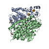 6rucC  5jshS S: Starting model for refinement C: citing same article ( |
|---|---|
| Similar structure data |


-Links
 PDBj
PDBj


- Assembly
| Deposited unit | 
| ||||||||||
|---|---|---|---|---|---|---|---|---|---|---|---|
| 1 |
| ||||||||||
| Unit cell |
|
-Components
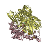
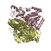
-Periplasmic [NiFeSe] hydrogenase, ... , 2 types, 2 molecules AB
| #1: Protein | Mass: 30261.568 Da / Num. of mol.: 1 Source method: isolated from a genetically manipulated source Source: (gene. exp.) Desulfovibrio vulgaris (strain Hildenborough / ATCC 29579 / DSM 644 / NCIMB 8303) (bacteria)Gene: hysB, DVU_1917 Production host: Desulfovibrio vulgaris str. Hildenborough (bacteria)References: UniProt: Q72AS4, ferredoxin hydrogenase |
|---|---|
| #2: Protein | Mass: 54469.246 Da / Num. of mol.: 1 / Mutation: G50T Source method: isolated from a genetically manipulated source Details: Cys 75 partially oxidized to sulfenate/sulfinate Source: (gene. exp.) Desulfovibrio vulgaris (strain Hildenborough / ATCC 29579 / DSM 644 / NCIMB 8303) (bacteria)Gene: hysA, DVU_1918 Production host: Desulfovibrio vulgaris str. Hildenborough (bacteria)References: UniProt: Q72AS3, ferredoxin hydrogenase |
-Non-polymers , 9 types, 811 molecules 
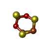

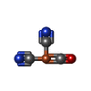


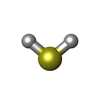


| #3: Chemical | Iron–sulfur cluster Mass: 351.640 Da / Num. of mol.: 3 / Source method: obtained synthetically / Formula: Fe4S4 Mass: 351.640 Da / Num. of mol.: 3 / Source method: obtained synthetically / Formula: Fe4S4#4: Chemical | ChemComp-6ML / |  Mass: 383.639 Da / Num. of mol.: 1 / Source method: obtained synthetically / Formula: Fe4O2S4 Mass: 383.639 Da / Num. of mol.: 1 / Source method: obtained synthetically / Formula: Fe4O2S4#5: Chemical | Glycerol Mass: 92.094 Da / Num. of mol.: 3 / Source method: obtained synthetically / Formula: C3H8O3 Mass: 92.094 Da / Num. of mol.: 3 / Source method: obtained synthetically / Formula: C3H8O3#6: Chemical | ChemComp-FCO / |  Mass: 135.890 Da / Num. of mol.: 1 / Source method: obtained synthetically / Formula: C3FeN2O Mass: 135.890 Da / Num. of mol.: 1 / Source method: obtained synthetically / Formula: C3FeN2O#7: Chemical | ChemComp-NI / | Nickel Mass: 58.693 Da / Num. of mol.: 1 / Source method: obtained synthetically / Formula: Ni Mass: 58.693 Da / Num. of mol.: 1 / Source method: obtained synthetically / Formula: Ni#8: Chemical | ChemComp-FE2 / |  Mass: 55.845 Da / Num. of mol.: 1 / Source method: obtained synthetically / Formula: Fe Mass: 55.845 Da / Num. of mol.: 1 / Source method: obtained synthetically / Formula: Fe#9: Chemical | ChemComp-H2S / | Hydrogen sulfide Mass: 34.081 Da / Num. of mol.: 1 / Source method: obtained synthetically / Formula: H2S Mass: 34.081 Da / Num. of mol.: 1 / Source method: obtained synthetically / Formula: H2S#10: Chemical | ChemComp-CL / | Chloride Mass: 35.453 Da / Num. of mol.: 1 / Source method: obtained synthetically / Formula: Cl Mass: 35.453 Da / Num. of mol.: 1 / Source method: obtained synthetically / Formula: Cl#11: Water | ChemComp-HOH / | WaterMass: 18.015 Da / Num. of mol.: 799 / Source method: isolated from a natural source / Formula: H2O |
|---|
-Experimental details
-Experiment
| Experiment | Method: X-RAY DIFFRACTION / Number of used crystals: 1 |
|---|
- Sample preparation
| Crystal | Density Matthews: 2.12 Å3/Da / Density % sol: 41.93 % / Description: Plate |
|---|---|
| Crystal grow | Temperature: 295 K / Method: vapor diffusion, sitting drop / pH: 7 / Details: 20% PEG 1500 (w/v), 0.1 M Tris-HCl pH 7 |
-Data collection
| Diffraction | Mean temperature: 100 K / Serial crystal experiment: N | ||||||||||||||||||||||||||||||
|---|---|---|---|---|---|---|---|---|---|---|---|---|---|---|---|---|---|---|---|---|---|---|---|---|---|---|---|---|---|---|---|
| Diffraction source | Source: SYNCHROTRON / Site: ESRF  / Beamline: ID29 / Wavelength: 0.8856 Å / Beamline: ID29 / Wavelength: 0.8856 Å | ||||||||||||||||||||||||||||||
| Detector | Type: DECTRIS PILATUS3 6M / Detector: PIXEL / Date: Dec 13, 2014 | ||||||||||||||||||||||||||||||
| Radiation | Protocol: SINGLE WAVELENGTH / Monochromatic (M) / Laue (L): M / Scattering type: x-ray | ||||||||||||||||||||||||||||||
| Radiation wavelength | Wavelength: 0.8856 Å / Relative weight: 1 | ||||||||||||||||||||||||||||||
| Reflection | Resolution: 1.1→45.38 Å / Num. obs: 263501 / % possible obs: 93.3 % / Redundancy: 2.3 % / Biso Wilson estimate: 11.03 Å2 / CC1/2: 0.994 / Rmerge(I) obs: 0.076 / Rpim(I) all: 0.061 / Rrim(I) all: 0.098 / Net I/σ(I): 6.1 / Num. measured all: 617436 | ||||||||||||||||||||||||||||||
| Reflection shell | Diffraction-ID: 1
|
-Phasing
| Phasing | Method: molecular replacement | |||||||||
|---|---|---|---|---|---|---|---|---|---|---|
| Phasing MR | Model details: Phaser MODE: MR_AUTO
|
- Processing
| Software |
| |||||||||||||||||||||||||||||||||||||||||||||||||||||||||||||||||||||||||||||||||||||||||||||||||||||||||||||||||||||||||||||||||||||||||||||||||||||||||||||||||||||||||||||||||||||||||||||||||||||||||||||||||||||||||
|---|---|---|---|---|---|---|---|---|---|---|---|---|---|---|---|---|---|---|---|---|---|---|---|---|---|---|---|---|---|---|---|---|---|---|---|---|---|---|---|---|---|---|---|---|---|---|---|---|---|---|---|---|---|---|---|---|---|---|---|---|---|---|---|---|---|---|---|---|---|---|---|---|---|---|---|---|---|---|---|---|---|---|---|---|---|---|---|---|---|---|---|---|---|---|---|---|---|---|---|---|---|---|---|---|---|---|---|---|---|---|---|---|---|---|---|---|---|---|---|---|---|---|---|---|---|---|---|---|---|---|---|---|---|---|---|---|---|---|---|---|---|---|---|---|---|---|---|---|---|---|---|---|---|---|---|---|---|---|---|---|---|---|---|---|---|---|---|---|---|---|---|---|---|---|---|---|---|---|---|---|---|---|---|---|---|---|---|---|---|---|---|---|---|---|---|---|---|---|---|---|---|---|---|---|---|---|---|---|---|---|---|---|---|---|---|---|---|---|
| Refinement | Method to determine structure: MOLECULAR REPLACEMENT Starting model: 5JSH Resolution: 1.1→45.38 Å / SU ML: 0.1206 / Cross valid method: THROUGHOUT / σ(F): 0.01 / Phase error: 18.2096 Stereochemistry target values: GeoStd + Monomer Library + CDL v1.2
| |||||||||||||||||||||||||||||||||||||||||||||||||||||||||||||||||||||||||||||||||||||||||||||||||||||||||||||||||||||||||||||||||||||||||||||||||||||||||||||||||||||||||||||||||||||||||||||||||||||||||||||||||||||||||
| Solvent computation | Shrinkage radii: 0.9 Å / VDW probe radii: 1.11 Å / Solvent model: FLAT BULK SOLVENT MODEL | |||||||||||||||||||||||||||||||||||||||||||||||||||||||||||||||||||||||||||||||||||||||||||||||||||||||||||||||||||||||||||||||||||||||||||||||||||||||||||||||||||||||||||||||||||||||||||||||||||||||||||||||||||||||||
| Displacement parameters | Biso mean: 15.24 Å2 | |||||||||||||||||||||||||||||||||||||||||||||||||||||||||||||||||||||||||||||||||||||||||||||||||||||||||||||||||||||||||||||||||||||||||||||||||||||||||||||||||||||||||||||||||||||||||||||||||||||||||||||||||||||||||
| Refinement step | Cycle: LAST / Resolution: 1.1→45.38 Å
| |||||||||||||||||||||||||||||||||||||||||||||||||||||||||||||||||||||||||||||||||||||||||||||||||||||||||||||||||||||||||||||||||||||||||||||||||||||||||||||||||||||||||||||||||||||||||||||||||||||||||||||||||||||||||
| Refine LS restraints |
| |||||||||||||||||||||||||||||||||||||||||||||||||||||||||||||||||||||||||||||||||||||||||||||||||||||||||||||||||||||||||||||||||||||||||||||||||||||||||||||||||||||||||||||||||||||||||||||||||||||||||||||||||||||||||
| LS refinement shell |
|
