 Movie
Movie Controller

[English] 日本語
 Yorodumi
- PDB-6rps: X-ray crystal structure of carbonic anhydrase XII complexed with ... -
+ Open data

- Basic information
| Entry | Database: PDB / ID: 6rps | ||||||
|---|---|---|---|---|---|---|---|
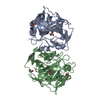
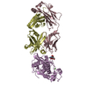
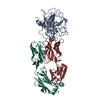
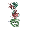
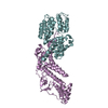
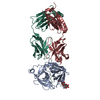
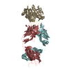
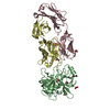
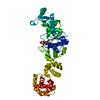
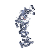
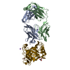
| Title | X-ray crystal structure of carbonic anhydrase XII complexed with a theranostic monoclonal antibody fragment | ||||||
 Components Components |
| ||||||
 Keywords Keywords |  LYASE / Anticancer drugs / Carbonic Anhydrase XII / complex / monoclonal antibody LYASE / Anticancer drugs / Carbonic Anhydrase XII / complex / monoclonal antibody | ||||||
| Function / homology |  Function and homology information Function and homology informationchloride ion homeostasis / estrous cycle / Reversible hydration of carbon dioxide / carbonic anhydrase / carbonate dehydratase activity / one-carbon metabolic process / basolateral plasma membrane / apical plasma membrane / zinc ion binding / membrane / plasma membraneSimilarity search - Function | ||||||
| Biological species |  Homo sapiens (human) Homo sapiens (human) | ||||||
| Method | X-RAY DIFFRACTION / MOLECULAR REPLACEMENT / Resolution: 2.79 Å | ||||||
 Authors Authors | Alterio, V. / Esposito, D. / De Simone, G. | ||||||
 Citation Citation | Journal: J.Mol.Biol. / Year: 2019 Title: Biochemical and Structural Insights into Carbonic Anhydrase XII/Fab6A10 Complex. Authors: Alterio, V. / Kellner, M. / Esposito, D. / Liesche-Starnecker, F. / Bua, S. / Supuran, C.T. / Monti, S.M. / Zeidler, R. / De Simone, G. | ||||||
| History |
|
- Structure visualization
| Structure viewer | Molecule: MolmilJmol/JSmol |
|---|
- Downloads & links
-Download
| PDBx/mmCIF format | 6rps.cif.gz | 279.5 KB | Display | PDBx/mmCIF format |
|---|---|---|---|---|
| PDB format | pdb6rps.ent.gz | 223.5 KB | Display | PDB format |
| PDBx/mmJSON format | 6rps.json.gz | Tree view | PDBx/mmJSON format | |
| Others |  Other downloads Other downloads |
-Validation report
| Arichive directory | https://data.pdbj.org/pub/pdb/validation_reports/rp/6rpsftp://data.pdbj.org/pub/pdb/validation_reports/rp/6rps | HTTPS FTP |
|---|
-Related structure data


-Links
 PDBj
PDBj

- Assembly
| Deposited unit | 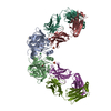
| ||||||||
|---|---|---|---|---|---|---|---|---|---|
| 1 | 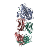
| ||||||||
| 2 | 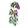
| ||||||||
| Unit cell |
|
-Components
-Protein , 1 types, 2 molecules AB
| #1: Protein | / Carbonate dehydratase XII / Carbonic anhydrase XII / CA-XII / Tumor antigen HOM-RCC-3.1.3 Mass: 31604.959 Da / Num. of mol.: 2 Source method: isolated from a genetically manipulated source Source: (gene. exp.) Homo sapiens (human) / Gene: CA12 / Production host:  Escherichia coli BL21(DE3) (bacteria) / Variant (production host): Rosetta / References: UniProt: O43570, carbonic anhydrase Escherichia coli BL21(DE3) (bacteria) / Variant (production host): Rosetta / References: UniProt: O43570, carbonic anhydrase |
|---|
-Antibody , 2 types, 4 molecules MLNH
| #2: Antibody | Fragment antigen-binding Mass: 23573.145 Da / Num. of mol.: 2 Source method: isolated from a genetically manipulated source Source: (gene. exp.) Homo sapiens (human) / Production host:   Cricetulus griseus (Chinese hamster) Cricetulus griseus (Chinese hamster)#3: Antibody | Fragment antigen-bindingMass: 24663.516 Da / Num. of mol.: 2 Source method: isolated from a genetically manipulated source Source: (gene. exp.) Homo sapiens (human) / Production host: Cricetulus griseus (Chinese hamster) |
|---|
-Non-polymers , 6 types, 34 molecules 


| #4: Chemical |  Mass: 65.409 Da / Num. of mol.: 2 / Source method: obtained synthetically / Formula: Zn Mass: 65.409 Da / Num. of mol.: 2 / Source method: obtained synthetically / Formula: Zn#5: Chemical | Acetate Mass: 59.044 Da / Num. of mol.: 2 / Source method: obtained synthetically / Formula: C2H3O2 Mass: 59.044 Da / Num. of mol.: 2 / Source method: obtained synthetically / Formula: C2H3O2#6: Chemical | Sulfate Mass: 96.063 Da / Num. of mol.: 2 / Source method: obtained synthetically / Formula: SO4 Mass: 96.063 Da / Num. of mol.: 2 / Source method: obtained synthetically / Formula: SO4#7: Chemical | ChemComp-CD /  Mass: 112.411 Da / Num. of mol.: 4 / Source method: obtained synthetically / Formula: Cd Mass: 112.411 Da / Num. of mol.: 4 / Source method: obtained synthetically / Formula: Cd#8: Chemical | ChemComp-CL / | Chloride Mass: 35.453 Da / Num. of mol.: 1 / Source method: obtained synthetically / Formula: Cl Mass: 35.453 Da / Num. of mol.: 1 / Source method: obtained synthetically / Formula: Cl#9: Water | ChemComp-HOH / | WaterMass: 18.015 Da / Num. of mol.: 23 / Source method: isolated from a natural source / Formula: H2O |
|---|
-Experimental details
-Experiment
| Experiment | Method: X-RAY DIFFRACTION / Number of used crystals: 1 |
|---|
- Sample preparation
| Crystal | Density Matthews: 3.59 Å3/Da / Density % sol: 65.75 % |
|---|---|
| Crystal grow | Temperature: 293 K / Method: vapor diffusion, hanging drop / pH: 5.5 Details: 1.5M AMMONIUM SULFATE 0.1M SODIUM ACETATE 0.02M CADMIUM CHLORIDE |
-Data collection
| Diffraction | Mean temperature: 100 K / Serial crystal experiment: N | |||||||||||||||||||||||||||||||||||||||||||||||||||||||||||||||||||||||||||||||||||||||||||||||||||||||||||||||||||||||||||||||||||||||||||||||||||||||||||||||||||||||||||||||||||||||||||||
|---|---|---|---|---|---|---|---|---|---|---|---|---|---|---|---|---|---|---|---|---|---|---|---|---|---|---|---|---|---|---|---|---|---|---|---|---|---|---|---|---|---|---|---|---|---|---|---|---|---|---|---|---|---|---|---|---|---|---|---|---|---|---|---|---|---|---|---|---|---|---|---|---|---|---|---|---|---|---|---|---|---|---|---|---|---|---|---|---|---|---|---|---|---|---|---|---|---|---|---|---|---|---|---|---|---|---|---|---|---|---|---|---|---|---|---|---|---|---|---|---|---|---|---|---|---|---|---|---|---|---|---|---|---|---|---|---|---|---|---|---|---|---|---|---|---|---|---|---|---|---|---|---|---|---|---|---|---|---|---|---|---|---|---|---|---|---|---|---|---|---|---|---|---|---|---|---|---|---|---|---|---|---|---|---|---|---|---|---|---|---|
| Diffraction source | Source: ROTATING ANODE / Type: RIGAKU MICROMAX-007 HF / Wavelength: 1.54178 Å | |||||||||||||||||||||||||||||||||||||||||||||||||||||||||||||||||||||||||||||||||||||||||||||||||||||||||||||||||||||||||||||||||||||||||||||||||||||||||||||||||||||||||||||||||||||||||||||
| Detector | Type: RIGAKU SATURN 944 / Detector: CCD / Date: Nov 15, 2017 | |||||||||||||||||||||||||||||||||||||||||||||||||||||||||||||||||||||||||||||||||||||||||||||||||||||||||||||||||||||||||||||||||||||||||||||||||||||||||||||||||||||||||||||||||||||||||||||
| Radiation | Protocol: SINGLE WAVELENGTH / Monochromatic (M) / Laue (L): M / Scattering type: x-ray | |||||||||||||||||||||||||||||||||||||||||||||||||||||||||||||||||||||||||||||||||||||||||||||||||||||||||||||||||||||||||||||||||||||||||||||||||||||||||||||||||||||||||||||||||||||||||||||
| Radiation wavelength | Wavelength: 1.54178 Å / Relative weight: 1 | |||||||||||||||||||||||||||||||||||||||||||||||||||||||||||||||||||||||||||||||||||||||||||||||||||||||||||||||||||||||||||||||||||||||||||||||||||||||||||||||||||||||||||||||||||||||||||||
| Reflection | Resolution: 2.79→50 Å / Num. obs: 56400 / % possible obs: 98.1 % / Redundancy: 4.8 % / Rmerge(I) obs: 0.091 / Rpim(I) all: 0.044 / Rrim(I) all: 0.102 / Χ2: 0.984 / Net I/σ(I): 8.6 / Num. measured all: 272450 | |||||||||||||||||||||||||||||||||||||||||||||||||||||||||||||||||||||||||||||||||||||||||||||||||||||||||||||||||||||||||||||||||||||||||||||||||||||||||||||||||||||||||||||||||||||||||||||
| Reflection shell | Diffraction-ID: 1
|
- Processing
| Software |
| ||||||||||||||||||||||||
|---|---|---|---|---|---|---|---|---|---|---|---|---|---|---|---|---|---|---|---|---|---|---|---|---|---|
| Refinement | Method to determine structure: MOLECULAR REPLACEMENT Starting model: 4WW8, 3FO0 Resolution: 2.79→49.65 Å / Cor.coef. Fo:Fc: 0.915 / Cor.coef. Fo:Fc free: 0.889 / SU B: 11.902 / SU ML: 0.222 / Cross valid method: THROUGHOUT / σ(F): 0 / ESU R: 0.645 / ESU R Free: 0.303 / Stereochemistry target values: MAXIMUM LIKELIHOOD / Details: U VALUES : REFINED INDIVIDUALLY
| ||||||||||||||||||||||||
| Solvent computation | Ion probe radii: 0.7 Å / Shrinkage radii: 0.7 Å / VDW probe radii: 1.1 Å / Solvent model: MASK | ||||||||||||||||||||||||
| Displacement parameters | Biso max: 116.98 Å2 / Biso mean: 36.415 Å2 / Biso min: 10.09 Å2
| ||||||||||||||||||||||||
| Refinement step | Cycle: final / Resolution: 2.79→49.65 Å
| ||||||||||||||||||||||||
| Refine LS restraints |
| ||||||||||||||||||||||||
| LS refinement shell | Resolution: 2.792→2.864 Å / Rfactor Rfree error: 0 / Total num. of bins used: 20
|
