 Movie
Movie Controller


+ Open data

- Basic information
| Entry | Database: PDB / ID: 6ro9 | ||||||
|---|---|---|---|---|---|---|---|
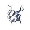
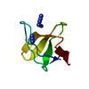
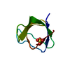
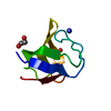
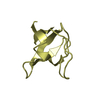
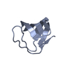
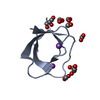
| Title | Human spectrin SH3 domain D48G, E7V, K60V | ||||||
 Components Components | Spectrin alpha, non-erythrocytic 1 | ||||||
 Keywords Keywords |  PROTEIN BINDING / Binding Domain PROTEIN BINDING / Binding Domain | ||||||
| Function / homology |  Function and homology information Function and homology informationclustering of voltage-gated sodium channels / paranode region of axon / node of Ranvier / actin filament capping / actin binding / cell cortex / cytoskeleton / calmodulin binding / calcium ion bindingSimilarity search - Function | ||||||
| Biological species |  Mola mola (ocean sunfish) Mola mola (ocean sunfish) | ||||||
| Method | X-RAY DIFFRACTION / SYNCHROTRON / MOLECULAR REPLACEMENT / Resolution: 1.814 Å | ||||||
 Authors Authors | Reverter, D. / Navarro, S. / Ventura, S. | ||||||
| Funding support |  Spain, 1items Spain, 1items
| ||||||
 Citation Citation | Journal: To Be Published Title: Human spectrin SH3 domain D48G, E7V, K60V Authors: Reverter, D. / Navarro, S. / Ventura, S. | ||||||
| History |
|
- Structure visualization
| Structure viewer | Molecule: MolmilJmol/JSmol |
|---|
- Downloads & links
-Download
| PDBx/mmCIF format | 6ro9.cif.gz | 36.6 KB | Display | PDBx/mmCIF format |
|---|---|---|---|---|
| PDB format | pdb6ro9.ent.gz | 23.8 KB | Display | PDB format |
| PDBx/mmJSON format | 6ro9.json.gz | Tree view | PDBx/mmJSON format | |
| Others |  Other downloads Other downloads |
-Validation report
| Arichive directory | https://data.pdbj.org/pub/pdb/validation_reports/ro/6ro9ftp://data.pdbj.org/pub/pdb/validation_reports/ro/6ro9 | HTTPS FTP |
|---|
-Related structure data
| Related structure data | 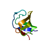 5fw9S S: Starting model for refinement |
|---|---|
| Similar structure data |


-Links
 PDBj
PDBj- Assembly
| Deposited unit | 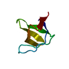
| ||||||||
|---|---|---|---|---|---|---|---|---|---|
| 1 |
| ||||||||
| Unit cell |
|
-Components
| #1: Protein | Mass: 6996.088 Da / Num. of mol.: 1 Source method: isolated from a genetically manipulated source Source: (gene. exp.) Mola mola (ocean sunfish) / Production host:  Escherichia coli (E. coli) / References: UniProt: A0A3Q3W9R3 Escherichia coli (E. coli) / References: UniProt: A0A3Q3W9R3 |
|---|---|
| #2: Water | ChemComp-HOH / Water Mass: 18.015 Da / Num. of mol.: 32 / Source method: isolated from a natural source / Formula: H2O Mass: 18.015 Da / Num. of mol.: 32 / Source method: isolated from a natural source / Formula: H2O |
-Experimental details
-Experiment
| Experiment | Method: X-RAY DIFFRACTION / Number of used crystals: 1 |
|---|
- Sample preparation
| Crystal | Density Matthews: 2.6 Å3/Da / Density % sol: 52.65 % |
|---|---|
| Crystal grow | Temperature: 316 K / Method: vapor diffusion, hanging drop / Details: 2M ammonium sulfate and 0.1M MES pH 6.5 |
-Data collection
| Diffraction | Mean temperature: 100 K / Serial crystal experiment: N |
|---|---|
| Diffraction source | Source: SYNCHROTRON / Site: ALBA / Beamline: XALOC / Wavelength: 0.97946 Å |
| Detector | Type: DECTRIS PILATUS 6M / Detector: PIXEL / Date: May 12, 2014 |
| Radiation | Protocol: SINGLE WAVELENGTH / Monochromatic (M) / Laue (L): M / Scattering type: x-ray |
| Radiation wavelength | Wavelength: 0.97946 Å / Relative weight: 1 |
| Reflection | Resolution: 1.814→41.819 Å / Num. obs: 6486 / % possible obs: 100 % / Redundancy: 5.9 % / CC1/2: 0.996 / Rmerge(I) obs: 0.116 / Rpim(I) all: 0.051 / Net I/σ(I): 11.4 |
| Reflection shell | Resolution: 1.814→1.82 Å / Rmerge(I) obs: 1.081 / Num. unique obs: 66 / CC1/2: 0.755 / Rpim(I) all: 0.477 |
- Processing
| Software |
| ||||||||||||||||||||||||||||||||||||||||
|---|---|---|---|---|---|---|---|---|---|---|---|---|---|---|---|---|---|---|---|---|---|---|---|---|---|---|---|---|---|---|---|---|---|---|---|---|---|---|---|---|---|
| Refinement | Method to determine structure: MOLECULAR REPLACEMENT Starting model: 5FW9 Resolution: 1.814→31.754 Å / SU ML: 0.16 / Cross valid method: FREE R-VALUE / σ(F): 1.34 / Phase error: 28.53
| ||||||||||||||||||||||||||||||||||||||||
| Solvent computation | Shrinkage radii: 0.9 Å / VDW probe radii: 1.11 Å | ||||||||||||||||||||||||||||||||||||||||
| Refinement step | Cycle: LAST / Resolution: 1.814→31.754 Å
| ||||||||||||||||||||||||||||||||||||||||
| Refine LS restraints |
| ||||||||||||||||||||||||||||||||||||||||
| LS refinement shell |
| ||||||||||||||||||||||||||||||||||||||||
| Refinement TLS params. | Method: refined / Origin x: -0.2336 Å / Origin y: 5.0522 Å / Origin z: 16.5741 Å
| ||||||||||||||||||||||||||||||||||||||||
| Refinement TLS group | Selection details: all |
