 Movie
Movie Controller


+ Open data

- Basic information
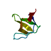
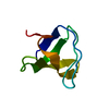
| Entry | Database: PDB / ID: 1u06 | ||||||
|---|---|---|---|---|---|---|---|
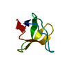
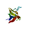
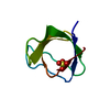
| Title | crystal structure of chicken alpha-spectrin SH3 domain | ||||||
 Components Components | Spectrin alpha chain, brain | ||||||
 Keywords Keywords |  STRUCTURAL PROTEIN / SH3 domain / beta barrel / five antiparallel beta sheets STRUCTURAL PROTEIN / SH3 domain / beta barrel / five antiparallel beta sheets | ||||||
| Function / homology |  Function and homology information Function and homology informationactin filament capping / costamere / cortical actin cytoskeleton / cell projection / actin filament binding / cell junction / actin cytoskeleton organization / calmodulin binding / calcium ion binding / plasma membraneSimilarity search - Function | ||||||
| Biological species |  Gallus gallus (chicken) Gallus gallus (chicken) | ||||||
| Method | X-RAY DIFFRACTION / SYNCHROTRON / MOLECULAR REPLACEMENT / Resolution: 1.49 Å | ||||||
 Authors Authors | Chevelkov, V. / Faelber, K. / Diehl, A. / Heinemann, U. / Oschkinat, H. / Reif, B. | ||||||
 Citation Citation | Journal: J.Biomol.Nmr / Year: 2005 Title: Detection of dynamic water molecules in a microcrystalline sample of the SH3 domain of alpha-spectrin by MAS solid-state NMR. Authors: Chevelkov, V. / Faelber, K. / Diehl, A. / Heinemann, U. / Oschkinat, H. / Reif, B. | ||||||
| History |
|
- Structure visualization
| Structure viewer | Molecule: MolmilJmol/JSmol |
|---|
- Downloads & links
-Download
| PDBx/mmCIF format | 1u06.cif.gz | 24.8 KB | Display | PDBx/mmCIF format |
|---|---|---|---|---|
| PDB format | pdb1u06.ent.gz | 15.5 KB | Display | PDB format |
| PDBx/mmJSON format | 1u06.json.gz | Tree view | PDBx/mmJSON format | |
| Others |  Other downloads Other downloads |
-Validation report
| Arichive directory | https://data.pdbj.org/pub/pdb/validation_reports/u0/1u06ftp://data.pdbj.org/pub/pdb/validation_reports/u0/1u06 | HTTPS FTP |
|---|
-Related structure data
| Related structure data | 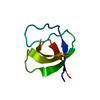 1shgS S: Starting model for refinement |
|---|---|
| Similar structure data |


-Links
 PDBj
PDBj- Assembly
| Deposited unit | 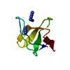
| ||||||||
|---|---|---|---|---|---|---|---|---|---|
| 1 |
| ||||||||
| Unit cell |
|
-Components
| #1: Protein | Mass: 7229.244 Da / Num. of mol.: 1 / Fragment: SH3 domain Source method: isolated from a genetically manipulated source Source: (gene. exp.) Gallus gallus (chicken) / Plasmid: pET3a / Species (production host): Escherichia coli / Production host:  Escherichia coli BL21(DE3) (bacteria) / Strain (production host): Bl21(DE3) / References: UniProt: P07751 Escherichia coli BL21(DE3) (bacteria) / Strain (production host): Bl21(DE3) / References: UniProt: P07751 | ||
|---|---|---|---|
| #2: Chemical | Azide  Mass: 42.020 Da / Num. of mol.: 3 / Source method: obtained synthetically / Formula: N3 Mass: 42.020 Da / Num. of mol.: 3 / Source method: obtained synthetically / Formula: N3#3: Water | ChemComp-HOH / | Water Mass: 18.015 Da / Num. of mol.: 53 / Source method: isolated from a natural source / Formula: H2O Mass: 18.015 Da / Num. of mol.: 53 / Source method: isolated from a natural source / Formula: H2O |
-Experimental details
-Experiment
| Experiment | Method: X-RAY DIFFRACTION / Number of used crystals: 1 |
|---|
- Sample preparation
| Crystal | Density Matthews: 2.52 Å3/Da / Density % sol: 50.7 % |
|---|---|
| Crystal grow | Temperature: 293 K / Method: vapor diffusion, hanging drop / pH: 7.2 Details: ammonium sulfate, pH 7.2, VAPOR DIFFUSION, HANGING DROP, temperature 293K |
-Data collection
| Diffraction | Mean temperature: 100 K |
|---|---|
| Diffraction source | Source: SYNCHROTRON / Site: SLS  / Beamline: X06SA / Wavelength: 1.006231 Å / Beamline: X06SA / Wavelength: 1.006231 Å |
| Detector | Type: MARRESEARCH / Detector: CCD / Date: Sep 17, 2003 / Details: mirrors |
| Radiation | Monochromator: SAGITALLY FOCUSED Si(111) / Protocol: SINGLE WAVELENGTH / Monochromatic (M) / Laue (L): M / Scattering type: x-ray |
| Radiation wavelength | Wavelength: 1.006231 Å / Relative weight: 1 |
| Reflection | Resolution: 1.49→40 Å / Num. all: 11423 / Num. obs: 11318 / % possible obs: 93.9 % / Observed criterion σ(F): 0 / Observed criterion σ(I): 0 / Redundancy: 6 % / Biso Wilson estimate: 23.3 Å2 / Rsym value: 0.038 / Net I/σ(I): 23.5 |
| Reflection shell | Resolution: 1.49→1.55 Å / Redundancy: 5.8 % / Mean I/σ(I) obs: 5.3 / Num. unique all: 1063 / Rsym value: 0.255 / % possible all: 79.9 |
- Processing
| Software |
| ||||||||||||||||||||
|---|---|---|---|---|---|---|---|---|---|---|---|---|---|---|---|---|---|---|---|---|---|
| Refinement | Method to determine structure: MOLECULAR REPLACEMENT Starting model: PDB ENTRY 1SHG Resolution: 1.49→32.11 Å Isotropic thermal model: OVERALL ANISOTROPIC B VALUE + TLS GROUP RESIDUE RANGE 7 - 61 Cross valid method: THROUGHOUT / σ(F): -3 / Stereochemistry target values: Engh & Huber
| ||||||||||||||||||||
| Displacement parameters | Biso mean: 14.615 Å2
| ||||||||||||||||||||
| Refinement step | Cycle: LAST / Resolution: 1.49→32.11 Å
| ||||||||||||||||||||
| Refine LS restraints |
| ||||||||||||||||||||
| LS refinement shell | Resolution: 1.49→1.529 Å /
|
