 Movie
Movie Controller


+ Open data

- Basic information
| Entry | Database: PDB / ID: 6rnj | ||||||
|---|---|---|---|---|---|---|---|
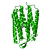
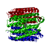
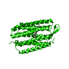
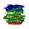
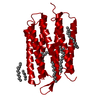
| Title | TR-SMX closed state structure (0-5ms) of bacteriorhodopsin | ||||||
 Components Components | Bacteriorhodopsin | ||||||
 Keywords Keywords | PROTON TRANSPORT / Retinal / time-resolved crystallography / serial crystallography / SMX | ||||||
| Function / homology |  Function and homology informationphotoreceptor activity / phototransduction / proton transmembrane transport / monoatomic ion channel activity / plasma membrane Function and homology informationphotoreceptor activity / phototransduction / proton transmembrane transport / monoatomic ion channel activity / plasma membraneSimilarity search - Function | ||||||
| Biological species |  Halobacterium salinarum NRC-1 (Halophile) Halobacterium salinarum NRC-1 (Halophile) | ||||||
| Method | X-RAY DIFFRACTION / SYNCHROTRON / MOLECULAR REPLACEMENT / molecular replacement / Resolution: 2.6 Å | ||||||
 Authors Authors | Weinert, T. / Skopintsev, P. / James, D. / Kekilli, D. / Furrer, F. / Bruenle, S. / Mous, S. / Nogly, P. / Standfuss, J. | ||||||
 Citation Citation | Journal: Science / Year: 2019 Title: Proton uptake mechanism in bacteriorhodopsin captured by serial synchrotron crystallography. Authors: Weinert, T. / Skopintsev, P. / James, D. / Dworkowski, F. / Panepucci, E. / Kekilli, D. / Furrer, A. / Brunle, S. / Mous, S. / Ozerov, D. / Nogly, P. / Wang, M. / Standfuss, J. | ||||||
| History |
|
- Structure visualization
| Structure viewer | Molecule: MolmilJmol/JSmol |
|---|
- Downloads & links
-Download
| PDBx/mmCIF format | 6rnj.cif.gz | 117.2 KB | Display | PDBx/mmCIF format |
|---|---|---|---|---|
| PDB format | pdb6rnj.ent.gz | 75.4 KB | Display | PDB format |
| PDBx/mmJSON format | 6rnj.json.gz | Tree view | PDBx/mmJSON format | |
| Others |  Other downloads Other downloads |
-Validation report
| Arichive directory | https://data.pdbj.org/pub/pdb/validation_reports/rn/6rnjftp://data.pdbj.org/pub/pdb/validation_reports/rn/6rnj | HTTPS FTP |
|---|
-Related structure data
| Related structure data |  6rphC  6rqoC  6rqpC  5b6zS S: Starting model for refinement C: citing same article ( |
|---|---|
| Similar structure data |
-Links
 PDBj
PDBj


- Assembly
| Deposited unit | 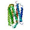
| ||||||||||
|---|---|---|---|---|---|---|---|---|---|---|---|
| 1 | 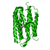
| ||||||||||
| Unit cell |
|
-Components
| #1: Protein | / BR / Bacterioopsin / BO Mass: 25061.615 Da / Num. of mol.: 1 / Source method: isolated from a natural source / Source: (natural) Halobacterium salinarum NRC-1 (Halophile) / References: UniProt: P02945 |
|---|---|
| #2: Chemical | ChemComp-RET / Retinal  Mass: 284.436 Da / Num. of mol.: 1 / Source method: obtained synthetically / Formula: C20H28O / Feature type: SUBJECT OF INVESTIGATION Mass: 284.436 Da / Num. of mol.: 1 / Source method: obtained synthetically / Formula: C20H28O / Feature type: SUBJECT OF INVESTIGATION |
| #3: Water | ChemComp-HOH / Water Mass: 18.015 Da / Num. of mol.: 8 / Source method: isolated from a natural source / Formula: H2O Mass: 18.015 Da / Num. of mol.: 8 / Source method: isolated from a natural source / Formula: H2O |
-Experimental details
-Experiment
| Experiment | Method: X-RAY DIFFRACTION / Number of used crystals: 1 |
|---|
- Sample preparation
| Crystal | Density Matthews: 2.44 Å3/Da / Density % sol: 49.63 % |
|---|---|
| Crystal grow | Temperature: 294 K / Method: lipidic cubic phase / pH: 5.6 / Details: 100 mM Na/K Phosphate buffer pH 5.6 30 % PEG 2000 |
-Data collection
| Diffraction |
| ||||||||||||||||||||||||||||||||||||
|---|---|---|---|---|---|---|---|---|---|---|---|---|---|---|---|---|---|---|---|---|---|---|---|---|---|---|---|---|---|---|---|---|---|---|---|---|---|
| Diffraction source |
| ||||||||||||||||||||||||||||||||||||
| Detector |
| ||||||||||||||||||||||||||||||||||||
| Radiation |
| ||||||||||||||||||||||||||||||||||||
| Radiation wavelength |
| ||||||||||||||||||||||||||||||||||||
| Reflection | Entry-ID: 6RNJ
| ||||||||||||||||||||||||||||||||||||
| Reflection shell |
| ||||||||||||||||||||||||||||||||||||
| Serial crystallography sample delivery |
| ||||||||||||||||||||||||||||||||||||
| Serial crystallography sample delivery injection |
|

-Phasing
| Phasing | Method: molecular replacement |
|---|
- Processing
| Software |
| ||||||||||||||||||||||||||||
|---|---|---|---|---|---|---|---|---|---|---|---|---|---|---|---|---|---|---|---|---|---|---|---|---|---|---|---|---|---|
| Refinement | Method to determine structure: MOLECULAR REPLACEMENT Starting model: 5B6Z Resolution: 2.6→38.47 Å / SU ML: 0.5373 / Cross valid method: THROUGHOUT / σ(F): 0.02 / Phase error: 44.8015
| ||||||||||||||||||||||||||||
| Solvent computation | Shrinkage radii: 0.9 Å / VDW probe radii: 1.11 Å | ||||||||||||||||||||||||||||
| Displacement parameters | Biso mean: 83.73 Å2 | ||||||||||||||||||||||||||||
| Refinement step | Cycle: LAST / Resolution: 2.6→38.47 Å
| ||||||||||||||||||||||||||||
| Refine LS restraints |
| ||||||||||||||||||||||||||||
| LS refinement shell |
|
