 Movie
Movie Controller

[English] 日本語
 Yorodumi
- PDB-6rmk: Bacteriorhodopsin, dark state, cell 2, refined using the same pro... -
+ Open data

- Basic information
| Entry | Database: PDB / ID: 6rmk | ||||||
|---|---|---|---|---|---|---|---|
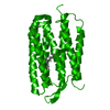
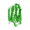
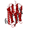
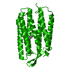
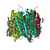
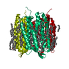
| Title | Bacteriorhodopsin, dark state, cell 2, refined using the same protocol as sub-ps time delays | ||||||
 Components Components | Bacteriorhodopsin | ||||||
 Keywords Keywords | MEMBRANE PROTEIN / proton pump / time-resolved crystallography / free-electron laser | ||||||
| Function / homology |  Function and homology informationphotoreceptor activity / phototransduction / proton transmembrane transport / monoatomic ion channel activity / plasma membrane Function and homology informationphotoreceptor activity / phototransduction / proton transmembrane transport / monoatomic ion channel activity / plasma membraneSimilarity search - Function | ||||||
| Biological species |  Halobacterium salinarum NRC-1 (Halophile) Halobacterium salinarum NRC-1 (Halophile) | ||||||
| Method | X-RAY DIFFRACTION / FREE ELECTRON LASER / MOLECULAR REPLACEMENT / Resolution: 1.8 Å | ||||||
 Authors Authors | Nass Kovacs, G. / Colletier, J.-P. / Gruenbein, M.L. / Stensitzki, T. / Batyuk, A. / Carbajo, S. / Doak, R.B. / Ehrenberg, D. / Foucar, L. / Gasper, R. ...Nass Kovacs, G. / Colletier, J.-P. / Gruenbein, M.L. / Stensitzki, T. / Batyuk, A. / Carbajo, S. / Doak, R.B. / Ehrenberg, D. / Foucar, L. / Gasper, R. / Gorel, A. / Hilpert, M. / Kloos, M. / Koglin, J. / Reinstein, J. / Roome, C.M. / Schlesinger, R. / Seaberg, M. / Shoeman, R.L. / Stricker, M. / Boutet, S. / Haacke, S. / Heberle, J. / Domratcheva, T. / Barends, T.R.M. / Schlichting, I. | ||||||
 Citation Citation | Journal: Nat Commun / Year: 2019 Title: Three-dimensional view of ultrafast dynamics in photoexcited bacteriorhodopsin. Authors: Nass Kovacs, G. / Colletier, J.P. / Grunbein, M.L. / Yang, Y. / Stensitzki, T. / Batyuk, A. / Carbajo, S. / Doak, R.B. / Ehrenberg, D. / Foucar, L. / Gasper, R. / Gorel, A. / Hilpert, M. / ...Authors: Nass Kovacs, G. / Colletier, J.P. / Grunbein, M.L. / Yang, Y. / Stensitzki, T. / Batyuk, A. / Carbajo, S. / Doak, R.B. / Ehrenberg, D. / Foucar, L. / Gasper, R. / Gorel, A. / Hilpert, M. / Kloos, M. / Koglin, J.E. / Reinstein, J. / Roome, C.M. / Schlesinger, R. / Seaberg, M. / Shoeman, R.L. / Stricker, M. / Boutet, S. / Haacke, S. / Heberle, J. / Heyne, K. / Domratcheva, T. / Barends, T.R.M. / Schlichting, I. | ||||||
| History |
|
- Structure visualization
| Structure viewer | Molecule: MolmilJmol/JSmol |
|---|
- Downloads & links
-Download
| PDBx/mmCIF format | 6rmk.cif.gz | 54.5 KB | Display | PDBx/mmCIF format |
|---|---|---|---|---|
| PDB format | pdb6rmk.ent.gz | 37.8 KB | Display | PDB format |
| PDBx/mmJSON format | 6rmk.json.gz | Tree view | PDBx/mmJSON format | |
| Others |  Other downloads Other downloads |
-Validation report
| Arichive directory | https://data.pdbj.org/pub/pdb/validation_reports/rm/6rmkftp://data.pdbj.org/pub/pdb/validation_reports/rm/6rmk | HTTPS FTP |
|---|
-Related structure data
| Related structure data | 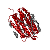 6ga1C  6ga2C  6ga3C  6ga4C  6ga5C 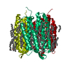 6ga6C  6ga7C  6ga8C  6ga9C 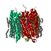 6gaaC  6gabC  6gacC  6gadC  6gaeC  6gafC  6gagC  6gahC  6gaiC 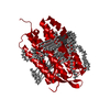 5b6vS S: Starting model for refinement C: citing same article ( |
|---|---|
| Similar structure data |


-Links
 PDBj
PDBj


- Assembly
| Deposited unit | 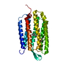
| ||||||||
|---|---|---|---|---|---|---|---|---|---|
| 1 | 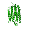
| ||||||||
| Unit cell |
|
-Components
| #1: Protein | / BR / Bacterioopsin / BO Mass: 25303.887 Da / Num. of mol.: 1 / Source method: isolated from a natural source / Source: (natural) Halobacterium salinarum NRC-1 / Variant: ATCC 700922 / JCM 11081 / NRC-1 / References: UniProt: P02945 |
|---|---|
| #2: Chemical | ChemComp-RET / Retinal  Mass: 284.436 Da / Num. of mol.: 1 / Source method: obtained synthetically / Formula: C20H28O Mass: 284.436 Da / Num. of mol.: 1 / Source method: obtained synthetically / Formula: C20H28O |
| #3: Water | ChemComp-HOH / Water Mass: 18.015 Da / Num. of mol.: 36 / Source method: isolated from a natural source / Formula: H2O Mass: 18.015 Da / Num. of mol.: 36 / Source method: isolated from a natural source / Formula: H2O |
-Experimental details
-Experiment
| Experiment | Method: X-RAY DIFFRACTION / Number of used crystals: 1 |
|---|
- Sample preparation
| Crystal | Density Matthews: 2.43 Å3/Da / Density % sol: 49.4 % |
|---|---|
| Crystal grow | Temperature: 293 K / Method: lipidic cubic phase Details: 32% (W/V) PEG 2000, 0.1 M K2HPO4 /NAH2PO4, IN HAMILTON SYRINGES, PH 5.6, LIPIDIC CUBIC PHASE, |
-Data collection
| Diffraction | Mean temperature: 293 K / Serial crystal experiment: N |
|---|---|
| Diffraction source | Source: FREE ELECTRON LASER / Site: SLAC LCLS  / Beamline: CXI / Wavelength: 1.26 Å / Beamline: CXI / Wavelength: 1.26 Å |
| Detector | Type: CS-PAD CXI-2 / Detector: PIXEL / Date: Aug 6, 2017 |
| Radiation | Protocol: SINGLE WAVELENGTH / Monochromatic (M) / Laue (L): M / Scattering type: x-ray |
| Radiation wavelength | Wavelength: 1.26 Å / Relative weight: 1 |
| Reflection | Resolution: 1.8→20.444 Å / Num. obs: 22403 / % possible obs: 100 % / Redundancy: 476 % / R split: 0.154 / Net I/σ(I): 9.9 |
| Reflection shell | Resolution: 1.8→1.85 Å / Num. unique obs: 1140 |
- Processing
| Software |
| |||||||||||||||||||||||||||||||||||||||||||||||||||||||||||||||
|---|---|---|---|---|---|---|---|---|---|---|---|---|---|---|---|---|---|---|---|---|---|---|---|---|---|---|---|---|---|---|---|---|---|---|---|---|---|---|---|---|---|---|---|---|---|---|---|---|---|---|---|---|---|---|---|---|---|---|---|---|---|---|---|---|
| Refinement | Method to determine structure: MOLECULAR REPLACEMENT Starting model: 5B6V Resolution: 1.8→20.441 Å / SU ML: 0.31 / Cross valid method: FREE R-VALUE / σ(F): 1.36 / Phase error: 28.33
| |||||||||||||||||||||||||||||||||||||||||||||||||||||||||||||||
| Solvent computation | Shrinkage radii: 0.9 Å / VDW probe radii: 1.11 Å | |||||||||||||||||||||||||||||||||||||||||||||||||||||||||||||||
| Refinement step | Cycle: LAST / Resolution: 1.8→20.441 Å
| |||||||||||||||||||||||||||||||||||||||||||||||||||||||||||||||
| Refine LS restraints |
| |||||||||||||||||||||||||||||||||||||||||||||||||||||||||||||||
| LS refinement shell |
|
