 Movie
Movie Controller

[English] 日本語
 Yorodumi
- PDB-6rmu: Crystal structure of disulphide-linked human C3d dimer in complex... -
+ Open data

- Basic information
| Entry | Database: PDB / ID: 6rmu | ||||||
|---|---|---|---|---|---|---|---|
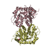
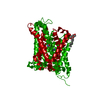
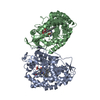
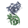
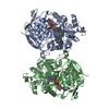
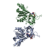
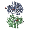
| Title | Crystal structure of disulphide-linked human C3d dimer in complex with Staphylococcus aureus complement subversion protein Sbi-IV | ||||||
 Components Components |
| ||||||
 Keywords Keywords |  IMMUNE SYSTEM / Complement / innate immunity / domain swap / Staphylococcus aureus IMMUNE SYSTEM / Complement / innate immunity / domain swap / Staphylococcus aureus | ||||||
| Function / homology |  Function and homology information Function and homology informationoviduct epithelium development / C5L2 anaphylatoxin chemotactic receptor binding / regulation of triglyceride biosynthetic process / positive regulation of activation of membrane attack complex / vertebrate eye-specific patterning / positive regulation of apoptotic cell clearance / complement-mediated synapse pruning / Alternative complement activation / positive regulation of lipid storage / positive regulation of G protein-coupled receptor signaling pathway ...oviduct epithelium development / C5L2 anaphylatoxin chemotactic receptor binding / regulation of triglyceride biosynthetic process / positive regulation of activation of membrane attack complex / vertebrate eye-specific patterning / positive regulation of apoptotic cell clearance / complement-mediated synapse pruning / Alternative complement activation / positive regulation of lipid storage / positive regulation of G protein-coupled receptor signaling pathway / positive regulation of phagocytosis, engulfment / complement receptor mediated signaling pathway / Activation of C3 and C5 / positive regulation of type IIa hypersensitivity / positive regulation of glucose transmembrane transport / complement-dependent cytotoxicity / complement activation, alternative pathway / complement activation / IgG binding / neuron remodeling / endopeptidase inhibitor activity / amyloid-beta clearance / positive regulation of vascular endothelial growth factor production / Purinergic signaling in leishmaniasis infection / complement activation, classical pathway / Peptide ligand-binding receptors / fatty acid metabolic process / Regulation of Complement cascade / response to bacterium / Post-translational protein phosphorylation / positive regulation of receptor-mediated endocytosis / positive regulation of angiogenesis / Regulation of Insulin-like Growth Factor (IGF) transport and uptake by Insulin-like Growth Factor Binding Proteins (IGFBPs) / azurophil granule lumen / Immunoregulatory interactions between a Lymphoid and a non-Lymphoid cell / G alpha (i) signalling events / secretory granule lumen / blood microparticle / immune response / inflammatory response / positive regulation of protein phosphorylation / G protein-coupled receptor signaling pathway / endoplasmic reticulum lumen / signaling receptor binding / Neutrophil degranulation / cell surface / signal transduction / protein-containing complex / extracellular space / extracellular exosome / extracellular region / plasma membraneSimilarity search - Function | ||||||
| Biological species |  Homo sapiens (human) Homo sapiens (human) Staphylococcus aureus subsp. aureus Mu50 (bacteria) Staphylococcus aureus subsp. aureus Mu50 (bacteria) | ||||||
| Method | X-RAY DIFFRACTION / SYNCHROTRON / MOLECULAR REPLACEMENT / Resolution: 2.4 Å | ||||||
 Authors Authors | Wahid, A.A. / van den Elsen, J.M.H. / Crennell, S.J. | ||||||
| Funding support |  United Kingdom, 1items United Kingdom, 1items
| ||||||
 Citation Citation | Journal: Front Immunol / Year: 2022 Title: Staphylococcal Complement Evasion Protein Sbi Stabilises C3d Dimers by Inducing an N-Terminal Helix Swap Authors: Dunphy, R.W. / Wahid, A.A. / Back, C.R. / Martin, R.L. / Watts, A.G. / Dodson, C.A. / Crennell, S.J. / van den Elsen, J.M.H. | ||||||
| History |
|
- Structure visualization
| Structure viewer | Molecule: MolmilJmol/JSmol |
|---|
- Downloads & links
-Download
| PDBx/mmCIF format | 6rmu.cif.gz | 283.8 KB | Display | PDBx/mmCIF format |
|---|---|---|---|---|
| PDB format | pdb6rmu.ent.gz | 232.5 KB | Display | PDB format |
| PDBx/mmJSON format | 6rmu.json.gz | Tree view | PDBx/mmJSON format | |
| Others |  Other downloads Other downloads |
-Validation report
| Arichive directory | https://data.pdbj.org/pub/pdb/validation_reports/rm/6rmuftp://data.pdbj.org/pub/pdb/validation_reports/rm/6rmu | HTTPS FTP |
|---|
-Related structure data
| Related structure data |  2wy7S S: Starting model for refinement |
|---|---|
| Similar structure data |


-Links
 PDBj
PDBj


- Assembly
| Deposited unit | 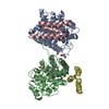
| ||||||||
|---|---|---|---|---|---|---|---|---|---|
| 1 |
| ||||||||
| Unit cell |
|
-Components
| #1: Protein | Complement component 3 / C3 and PZP-like alpha-2-macroglobulin domain-containing protein 1 Mass: 34778.812 Da / Num. of mol.: 2 Source method: isolated from a genetically manipulated source Source: (gene. exp.) Homo sapiens (human) / Gene: C3, CPAMD1 / Production host: Escherichia coli BL21(DE3) (bacteria) / References: UniProt: P01024#2: Protein | Mass: 9269.509 Da / Num. of mol.: 2 Source method: isolated from a genetically manipulated source Source: (gene. exp.) Staphylococcus aureus subsp. aureus Mu50 (bacteria)Gene: sbi, SAV2418 / Plasmid: pQE30 / Production host: Escherichia coli BL21(DE3) (bacteria) / References: UniProt: Q931F4#3: Chemical | ChemComp-PEG / | Diethylene glycol  Mass: 106.120 Da / Num. of mol.: 1 / Source method: obtained synthetically / Formula: C4H10O3 Mass: 106.120 Da / Num. of mol.: 1 / Source method: obtained synthetically / Formula: C4H10O3#4: Chemical | ChemComp-EDO / | Ethylene glycol  Mass: 62.068 Da / Num. of mol.: 1 / Source method: obtained synthetically / Formula: C2H6O2 Mass: 62.068 Da / Num. of mol.: 1 / Source method: obtained synthetically / Formula: C2H6O2#5: Water | ChemComp-HOH / | Water Mass: 18.015 Da / Num. of mol.: 259 / Source method: isolated from a natural source / Formula: H2O Mass: 18.015 Da / Num. of mol.: 259 / Source method: isolated from a natural source / Formula: H2O |
|---|
-Experimental details
-Experiment
| Experiment | Method: X-RAY DIFFRACTION / Number of used crystals: 1 |
|---|
- Sample preparation
| Crystal | Density Matthews: 2.86 Å3/Da / Density % sol: 56.96 % |
|---|---|
| Crystal grow | Temperature: 291 K / Method: vapor diffusion, hanging drop / pH: 8.07 Details: 0.2 M sodium citrate tribasic dihydrate, 20% PEG 3350. |
-Data collection
| Diffraction | Mean temperature: 100 K / Serial crystal experiment: N |
|---|---|
| Diffraction source | Source: SYNCHROTRON / Site: Diamond / Beamline: I04 / Wavelength: 0.9795 Å |
| Detector | Type: DECTRIS PILATUS 6M / Detector: PIXEL / Date: Jul 10, 2018 |
| Radiation | Protocol: SINGLE WAVELENGTH / Monochromatic (M) / Laue (L): M / Scattering type: x-ray |
| Radiation wavelength | Wavelength: 0.9795 Å / Relative weight: 1 |
| Reflection | Resolution: 2.4→118.93 Å / Num. obs: 40413 / % possible obs: 99.9 % / Redundancy: 5.7 % / CC1/2: 0.994 / Rmerge(I) obs: 0.132 / Rpim(I) all: 0.06 / Net I/σ(I): 8.5 |
| Reflection shell | Resolution: 2.4→2.49 Å / Redundancy: 5.8 % / Rmerge(I) obs: 0.587 / Mean I/σ(I) obs: 2.8 / Num. unique obs: 4153 / CC1/2: 0.791 / Rpim(I) all: 0.266 / % possible all: 99.1 |
- Processing
| Software |
| ||||||||||||||||||||||||||||||||||||||||||||||||||||||||||||||||||||||||||||||||||||||||||||||||||||||||||||||||
|---|---|---|---|---|---|---|---|---|---|---|---|---|---|---|---|---|---|---|---|---|---|---|---|---|---|---|---|---|---|---|---|---|---|---|---|---|---|---|---|---|---|---|---|---|---|---|---|---|---|---|---|---|---|---|---|---|---|---|---|---|---|---|---|---|---|---|---|---|---|---|---|---|---|---|---|---|---|---|---|---|---|---|---|---|---|---|---|---|---|---|---|---|---|---|---|---|---|---|---|---|---|---|---|---|---|---|---|---|---|---|---|---|---|
| Refinement | Method to determine structure: MOLECULAR REPLACEMENT Starting model: 2WY7 Resolution: 2.4→82.733 Å / SU ML: 0.25 / Cross valid method: FREE R-VALUE / σ(F): 1.46 / Phase error: 20.49
| ||||||||||||||||||||||||||||||||||||||||||||||||||||||||||||||||||||||||||||||||||||||||||||||||||||||||||||||||
| Solvent computation | Shrinkage radii: 0.9 Å / VDW probe radii: 1.11 Å | ||||||||||||||||||||||||||||||||||||||||||||||||||||||||||||||||||||||||||||||||||||||||||||||||||||||||||||||||
| Refinement step | Cycle: LAST / Resolution: 2.4→82.733 Å
| ||||||||||||||||||||||||||||||||||||||||||||||||||||||||||||||||||||||||||||||||||||||||||||||||||||||||||||||||
| Refine LS restraints |
| ||||||||||||||||||||||||||||||||||||||||||||||||||||||||||||||||||||||||||||||||||||||||||||||||||||||||||||||||
| LS refinement shell |
|
