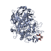
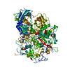
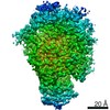
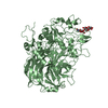
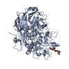
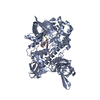
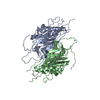
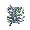
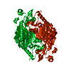
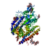
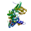
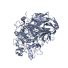
Journal: Nat Commun / Year: 2020 Title: Immunodominant proteins P1 and P40/P90 from human pathogen Mycoplasma pneumoniae. Authors: David Vizarraga / Akihiro Kawamoto / U Matsumoto / Ramiro Illanes / Rosa Pérez-Luque / Jesús Martín / Rocco Mazzolini / Paula Bierge / Oscar Q Pich / Mateu Espasa / Isabel Sanfeliu / ...Authors: David Vizarraga / Akihiro Kawamoto / U Matsumoto / Ramiro Illanes / Rosa Pérez-Luque / Jesús Martín / Rocco Mazzolini / Paula Bierge / Oscar Q Pich / Mateu Espasa / Isabel Sanfeliu / Juliana Esperalba / Miguel Fernández-Huerta / Margot P Scheffer / Jaume Pinyol / Achilleas S Frangakis / Maria Lluch-Senar / Shigetarou Mori / Keigo Shibayama / Tsuyoshi Kenri / Takayuki Kato / Keiichi Namba / Ignacio Fita / Makoto Miyata / David Aparicio / Abstract: Mycoplasma pneumoniae is a bacterial human pathogen that causes primary atypical pneumonia. M. pneumoniae motility and infectivity are mediated by the immunodominant proteins P1 and P40/P90, which ...Mycoplasma pneumoniae is a bacterial human pathogen that causes primary atypical pneumonia. M. pneumoniae motility and infectivity are mediated by the immunodominant proteins P1 and P40/P90, which form a transmembrane adhesion complex. Here we report the structure of P1, determined by X-ray crystallography and cryo-electron microscopy, and the X-ray structure of P40/P90. Contrary to what had been suggested, the binding site for sialic acid was found in P40/P90 and not in P1. Genetic and clinical variability concentrates on the N-terminal domain surfaces of P1 and P40/P90. Polyclonal antibodies generated against the mostly conserved C-terminal domain of P1 inhibited adhesion of M. pneumoniae, and serology assays with sera from infected patients were positive when tested against this C-terminal domain. P40/P90 also showed strong reactivity against human infected sera. The architectural elements determined for P1 and P40/P90 open new possibilities in vaccine development against M. pneumoniae infections.
In the structure databanks used in Yorodumi, some data are registered as the other names, "COVID-19 virus" and "2019-nCoV". Here are the details of the virus and the list of structure data.
Jan 31, 2019. EMDB accession codes are about to change! (news from PDBe EMDB page)
EMDB accession codes are about to change! (news from PDBe EMDB page)
The allocation of 4 digits for EMDB accession codes will soon come to an end. Whilst these codes will remain in use, new EMDB accession codes will include an additional digit and will expand incrementally as the available range of codes is exhausted. The current 4-digit format prefixed with “EMD-” (i.e. EMD-XXXX) will advance to a 5-digit format (i.e. EMD-XXXXX), and so on. It is currently estimated that the 4-digit codes will be depleted around Spring 2019, at which point the 5-digit format will come into force.
The EM Navigator/Yorodumi systems omit the EMD- prefix.
Related info.:Q: What is EMD? / ID/Accession-code notation in Yorodumi/EM Navigator
Yorodumi is a browser for structure data from EMDB, PDB, SASBDB, etc.
This page is also the successor to EM Navigator detail page, and also detail information page/front-end page for Omokage search.
The word "yorodu" (or yorozu) is an old Japanese word meaning "ten thousand". "mi" (miru) is to see.
Related info.:EMDB / PDB / SASBDB / Comparison of 3 databanks / Yorodumi Search / Aug 31, 2016. New EM Navigator & Yorodumi / Yorodumi Papers / Jmol/JSmol / Function and homology information / Changes in new EM Navigator and Yorodumi
 Movie
Movie Controller


 Open data
 Basic information
 Components
 Keywords
 CELL ADHESION /
  Function and homology information
 Mycoplasma pneumoniae (Filterable agent of primary atypical pneumonia)
 Authors
 Spain, 1items
  Citation

 Structure visualization
 Downloads & links
 Other downloads
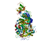


 PDBj
PDBj Assembly
 Mass: 18.015 Da / Num. of mol.: 64 / Source method: isolated from a natural source / Formula: H2O
 Sample preparation
 Processing