 Movie
Movie Controller


+ Open data

- Basic information
| Entry | Database: PDB / ID: 6r75 | |||||||||
|---|---|---|---|---|---|---|---|---|---|---|
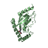
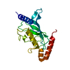
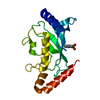
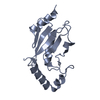
| Title | Crystal structure of human Ube2T E54R mutant | |||||||||
 Components Components | Ubiquitin-conjugating enzyme E2 T | |||||||||
 Keywords Keywords |  LIGASE / DNA repair / E2 / Ubiquitination / allostery LIGASE / DNA repair / E2 / Ubiquitination / allostery | |||||||||
| Function / homology |  Function and homology information Function and homology informationprotein K29-linked ubiquitination / protein K27-linked ubiquitination / protein K6-linked ubiquitination / protein K11-linked ubiquitination / E2 ubiquitin-conjugating enzyme / ubiquitin conjugating enzyme activity / protein monoubiquitination / protein K63-linked ubiquitination / protein K48-linked ubiquitination / protein autoubiquitination ...protein K29-linked ubiquitination / protein K27-linked ubiquitination / protein K6-linked ubiquitination / protein K11-linked ubiquitination / E2 ubiquitin-conjugating enzyme / ubiquitin conjugating enzyme activity / protein monoubiquitination / protein K63-linked ubiquitination / protein K48-linked ubiquitination / protein autoubiquitination / Synthesis of active ubiquitin: roles of E1 and E2 enzymes / Fanconi Anemia Pathway / protein polyubiquitination / ubiquitin-protein transferase activity / DNA repair / ubiquitin protein ligase binding / DNA damage response / chromatin binding / nucleolus / nucleoplasm / ATP binding / nucleusSimilarity search - Function | |||||||||
| Biological species |  Homo sapiens (human) Homo sapiens (human) | |||||||||
| Method | X-RAY DIFFRACTION / SYNCHROTRON / MOLECULAR REPLACEMENT / molecular replacement / Resolution: 2 Å | |||||||||
 Authors Authors | Chaugule, V.K. / Rennie, M.L. / Walden, H. / Arkinson, C. / Kamarainen, O. / Toth, R. | |||||||||
| Funding support |  United Kingdom, 2items United Kingdom, 2items
| |||||||||
 Citation Citation | Journal: Nat.Chem.Biol. / Year: 2020 Title: Allosteric mechanism for site-specific ubiquitination of FANCD2. Authors: Chaugule, V.K. / Arkinson, C. / Rennie, M.L. / Kamarainen, O. / Toth, R. / Walden, H. | |||||||||
| History |
|
- Structure visualization
| Structure viewer | Molecule: MolmilJmol/JSmol |
|---|
- Downloads & links
-Download
| PDBx/mmCIF format | 6r75.cif.gz | 47.4 KB | Display | PDBx/mmCIF format |
|---|---|---|---|---|
| PDB format | pdb6r75.ent.gz | 30.7 KB | Display | PDB format |
| PDBx/mmJSON format | 6r75.json.gz | Tree view | PDBx/mmJSON format | |
| Others |  Other downloads Other downloads |
-Validation report
| Arichive directory | https://data.pdbj.org/pub/pdb/validation_reports/r7/6r75ftp://data.pdbj.org/pub/pdb/validation_reports/r7/6r75 | HTTPS FTP |
|---|
-Related structure data
| Related structure data | 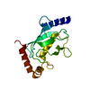 1yh2S S: Starting model for refinement |
|---|---|
| Similar structure data |


-Links
 PDBj
PDBj

- Assembly
| Deposited unit | 
| ||||||||
|---|---|---|---|---|---|---|---|---|---|
| 1 |
| ||||||||
| Unit cell |
|
-Components
| #1: Protein | Mass: 22880.250 Da / Num. of mol.: 1 / Mutation: E54R Source method: isolated from a genetically manipulated source Source: (gene. exp.) Homo sapiens (human) / Gene: UBE2T, HSPC150, PIG50 / Production host:  Escherichia coli (E. coli) Escherichia coli (E. coli)References: UniProt: Q9NPD8, E2 ubiquitin-conjugating enzyme |
|---|---|
| #2: Water | ChemComp-HOH / Water Mass: 18.015 Da / Num. of mol.: 57 / Source method: isolated from a natural source / Formula: H2O Mass: 18.015 Da / Num. of mol.: 57 / Source method: isolated from a natural source / Formula: H2O |
-Experimental details
-Experiment
| Experiment | Method: X-RAY DIFFRACTION / Number of used crystals: 1 |
|---|
- Sample preparation
| Crystal | Density Matthews: 2.21 Å3/Da / Density % sol: 44.35 % |
|---|---|
| Crystal grow | Temperature: 277 K / Method: vapor diffusion, sitting drop Details: 0.1 M Tris-HCl pH 8.5, 20% v/v glycerol ethoxylate, 3% v/v poly(ethylene imine) |
-Data collection
| Diffraction | Mean temperature: 100 K / Serial crystal experiment: N | ||||||||||||||||||||||||
|---|---|---|---|---|---|---|---|---|---|---|---|---|---|---|---|---|---|---|---|---|---|---|---|---|---|
| Diffraction source | Source: SYNCHROTRON / Site: ESRF  / Beamline: MASSIF-1 / Wavelength: 0.966 Å / Beamline: MASSIF-1 / Wavelength: 0.966 Å | ||||||||||||||||||||||||
| Detector | Type: DECTRIS PILATUS3 2M / Detector: PIXEL / Date: Jun 14, 2017 | ||||||||||||||||||||||||
| Radiation | Protocol: SINGLE WAVELENGTH / Monochromatic (M) / Laue (L): M / Scattering type: x-ray | ||||||||||||||||||||||||
| Radiation wavelength | Wavelength: 0.966 Å / Relative weight: 1 | ||||||||||||||||||||||||
| Reflection | Resolution: 2→46.19 Å / Num. obs: 9878 / % possible obs: 88.7 % / Redundancy: 3.5 % / CC1/2: 0.997 / Rmerge(I) obs: 0.045 / Rpim(I) all: 0.028 / Rrim(I) all: 0.053 / Net I/σ(I): 16.7 / Num. measured all: 34140 / Scaling rejects: 182 | ||||||||||||||||||||||||
| Reflection shell | Diffraction-ID: 1
|
-Phasing
| Phasing | Method: molecular replacement |
|---|
- Processing
| Software |
| ||||||||||||||||||||||||||||||||||||||||||||||||||||||||||||
|---|---|---|---|---|---|---|---|---|---|---|---|---|---|---|---|---|---|---|---|---|---|---|---|---|---|---|---|---|---|---|---|---|---|---|---|---|---|---|---|---|---|---|---|---|---|---|---|---|---|---|---|---|---|---|---|---|---|---|---|---|---|
| Refinement | Method to determine structure: MOLECULAR REPLACEMENT Starting model: 1yh2 Resolution: 2→39.21 Å / Cor.coef. Fo:Fc: 0.913 / Cor.coef. Fo:Fc free: 0.88 / SU B: 5.057 / SU ML: 0.137 / SU R Cruickshank DPI: 0.2717 / Cross valid method: THROUGHOUT / σ(F): 0 / ESU R: 0.272 / ESU R Free: 0.211 Details: HYDROGENS HAVE BEEN ADDED IN THE RIDING POSITIONS U VALUES : REFINED INDIVIDUALLY
| ||||||||||||||||||||||||||||||||||||||||||||||||||||||||||||
| Solvent computation | Ion probe radii: 0.8 Å / Shrinkage radii: 0.8 Å / VDW probe radii: 1.2 Å | ||||||||||||||||||||||||||||||||||||||||||||||||||||||||||||
| Displacement parameters | Biso max: 67.64 Å2 / Biso mean: 24.326 Å2 / Biso min: 14.21 Å2
| ||||||||||||||||||||||||||||||||||||||||||||||||||||||||||||
| Refinement step | Cycle: final / Resolution: 2→39.21 Å
| ||||||||||||||||||||||||||||||||||||||||||||||||||||||||||||
| Refine LS restraints |
| ||||||||||||||||||||||||||||||||||||||||||||||||||||||||||||
| LS refinement shell | Resolution: 2→2.052 Å / Rfactor Rfree error: 0 / Total num. of bins used: 20
|
