 Movie
Movie Controller

[English] 日本語
 Yorodumi
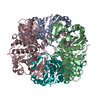
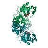
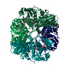
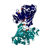
- PDB-6qun: Crystal structure of AtGapC1 with the catalytic Cys149 irreversib... -
+ Open data

- Basic information
| Entry | Database: PDB / ID: 6qun | ||||||
|---|---|---|---|---|---|---|---|
| Title | Crystal structure of AtGapC1 with the catalytic Cys149 irreversibly oxidized by H2O2 treatment | ||||||
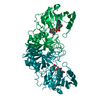
 Components Components | Glyceraldehyde-3-phosphate dehydrogenase GAPC1, cytosolic | ||||||
 Keywords Keywords |  OXIDOREDUCTASE / Rossman fold / NAD binding / Glycolytic process / oxidative stress / Hydrogen peroxide OXIDOREDUCTASE / Rossman fold / NAD binding / Glycolytic process / oxidative stress / Hydrogen peroxide | ||||||
| Function / homology |  Function and homology information Function and homology informationfruit development / glyceraldehyde-3-phosphate dehydrogenase (NADP+) (non-phosphorylating) activity / salicylic acid binding / seed development / response to sucrose / glyceraldehyde-3-phosphate dehydrogenase (phosphorylating) / plant-type vacuole / apoplast / glyceraldehyde-3-phosphate dehydrogenase (NAD+) (phosphorylating) activity / mitochondrial envelope ...fruit development / glyceraldehyde-3-phosphate dehydrogenase (NADP+) (non-phosphorylating) activity / salicylic acid binding / seed development / response to sucrose / glyceraldehyde-3-phosphate dehydrogenase (phosphorylating) / plant-type vacuole / apoplast / glyceraldehyde-3-phosphate dehydrogenase (NAD+) (phosphorylating) activity / mitochondrial envelope / response to redox state / chloroplast / gluconeogenesis / glycolytic process / response to hydrogen peroxide / NAD binding / NADP binding / response to heat / response to oxidative stress / copper ion binding / positive regulation of DNA-templated transcription / mitochondrion / DNA binding / nucleus / plasma membrane / cytosolSimilarity search - Function | ||||||
| Biological species |  Arabidopsis thaliana (thale cress) Arabidopsis thaliana (thale cress) | ||||||
| Method | X-RAY DIFFRACTION / SYNCHROTRON / MOLECULAR REPLACEMENT / Resolution: 3 Å | ||||||
 Authors Authors | Fermani, S. / Zaffagnini, M. / Falini, G. / Trost, P. | ||||||
| Funding support |  Italy, 1items Italy, 1items
| ||||||
 Citation Citation | Journal: Proc.Natl.Acad.Sci.USA / Year: 2019 Title: Glutathionylation primes soluble glyceraldehyde-3-phosphate dehydrogenase for late collapse into insoluble aggregates. Authors: Zaffagnini, M. / Marchand, C.H. / Malferrari, M. / Murail, S. / Bonacchi, S. / Genovese, D. / Montalti, M. / Venturoli, G. / Falini, G. / Baaden, M. / Lemaire, S.D. / Fermani, S. / Trost, P. | ||||||
| History |
|
- Structure visualization
| Structure viewer | Molecule: MolmilJmol/JSmol |
|---|
- Downloads & links
-Download
| PDBx/mmCIF format | 6qun.cif.gz | 277.8 KB | Display | PDBx/mmCIF format |
|---|---|---|---|---|
| PDB format | pdb6qun.ent.gz | 229.3 KB | Display | PDB format |
| PDBx/mmJSON format | 6qun.json.gz | Tree view | PDBx/mmJSON format | |
| Others |  Other downloads Other downloads |
-Validation report
| Arichive directory | https://data.pdbj.org/pub/pdb/validation_reports/qu/6qunftp://data.pdbj.org/pub/pdb/validation_reports/qu/6qun | HTTPS FTP |
|---|
-Related structure data
| Related structure data |  6quqC  4z0hS S: Starting model for refinement C: citing same article ( |
|---|---|
| Similar structure data |


-Links
 PDBj
PDBj
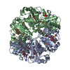
- Assembly
| Deposited unit | 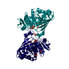
| ||||||||
|---|---|---|---|---|---|---|---|---|---|
| 1 | 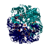
| ||||||||
| Unit cell |
| ||||||||
| Components on special symmetry positions |
|
-Components
| #1: Protein | Mass: 36549.707 Da / Num. of mol.: 2 Source method: isolated from a genetically manipulated source Details: The catalytic cysteine is oxidated to sulphinic acid Source: (gene. exp.) Arabidopsis thaliana (thale cress) / Gene: GAPC1, GAPC, GAPDH, At3g04120, T6K12.26 / Production host:  Escherichia coli (E. coli) Escherichia coli (E. coli)References: UniProt: P25858, glyceraldehyde-3-phosphate dehydrogenase (phosphorylating)#2: Chemical | Nicotinamide adenine dinucleotide  Mass: 663.425 Da / Num. of mol.: 2 / Source method: obtained synthetically / Formula: C21H27N7O14P2 / Comment: NAD*YM Mass: 663.425 Da / Num. of mol.: 2 / Source method: obtained synthetically / Formula: C21H27N7O14P2 / Comment: NAD*YM#3: Chemical | ChemComp-SO4 / Sulfate  Mass: 96.063 Da / Num. of mol.: 7 / Source method: obtained synthetically / Formula: SO4 Mass: 96.063 Da / Num. of mol.: 7 / Source method: obtained synthetically / Formula: SO4#4: Water | ChemComp-HOH / | Water Mass: 18.015 Da / Num. of mol.: 33 / Source method: isolated from a natural source / Formula: H2O Mass: 18.015 Da / Num. of mol.: 33 / Source method: isolated from a natural source / Formula: H2O |
|---|
-Experimental details
-Experiment
| Experiment | Method: X-RAY DIFFRACTION / Number of used crystals: 1 |
|---|
- Sample preparation
| Crystal | Density Matthews: 2.43 Å3/Da / Density % sol: 49.31 % |
|---|---|
| Crystal grow | Temperature: 293 K / Method: vapor diffusion, hanging drop / pH: 7.5 Details: 3.0 M (NH4)2SO4, 0.1 M Hepes-NaOH (pH 7.5), 0.1 mM H2O2 |
-Data collection
| Diffraction | Mean temperature: 100 K / Serial crystal experiment: N |
|---|---|
| Diffraction source | Source: SYNCHROTRON / Site: ELETTRA / Beamline: 5.2R / Wavelength: 1.26 Å |
| Detector | Type: DECTRIS PILATUS 2M / Detector: PIXEL / Date: May 15, 2012 Details: Cilindrical Mirror with 50 nm Pt-coating, oridal Mirros with 50 nm Pt-coating |
| Radiation | Monochromator: Si 111 double crystal / Protocol: SINGLE WAVELENGTH / Monochromatic (M) / Laue (L): M / Scattering type: x-ray |
| Radiation wavelength | Wavelength: 1.26 Å / Relative weight: 1 |
| Reflection | Resolution: 3→47.79 Å / Num. obs: 15798 / % possible obs: 99.8 % / Observed criterion σ(I): -3 / Redundancy: 7.8 % / Biso Wilson estimate: 62.2 Å2 / Rmerge(I) obs: 0.112 / Rpim(I) all: 0.043 / Rrim(I) all: 0.12 / Net I/σ(I): 13.5 |
| Reflection shell | Resolution: 3→3.16 Å / Redundancy: 8.1 % / Rmerge(I) obs: 0.453 / Mean I/σ(I) obs: 4.1 / Num. unique obs: 2164 / Rpim(I) all: 0.168 / Rrim(I) all: 0.483 / % possible all: 98.6 |
- Processing
| Software |
| |||||||||||||||||||||||||||||||||||||||||||||||||||||||||||||||||||||||||||||
|---|---|---|---|---|---|---|---|---|---|---|---|---|---|---|---|---|---|---|---|---|---|---|---|---|---|---|---|---|---|---|---|---|---|---|---|---|---|---|---|---|---|---|---|---|---|---|---|---|---|---|---|---|---|---|---|---|---|---|---|---|---|---|---|---|---|---|---|---|---|---|---|---|---|---|---|---|---|---|
| Refinement | Method to determine structure: MOLECULAR REPLACEMENT Starting model: 4Z0H Resolution: 3→47.785 Å / SU ML: 0.47 / Cross valid method: FREE R-VALUE / Phase error: 29.72
| |||||||||||||||||||||||||||||||||||||||||||||||||||||||||||||||||||||||||||||
| Solvent computation | Shrinkage radii: 0.9 Å / VDW probe radii: 1.11 Å | |||||||||||||||||||||||||||||||||||||||||||||||||||||||||||||||||||||||||||||
| Displacement parameters | Biso mean: 72.9 Å2 | |||||||||||||||||||||||||||||||||||||||||||||||||||||||||||||||||||||||||||||
| Refinement step | Cycle: LAST / Resolution: 3→47.785 Å
| |||||||||||||||||||||||||||||||||||||||||||||||||||||||||||||||||||||||||||||
| Refine LS restraints |
| |||||||||||||||||||||||||||||||||||||||||||||||||||||||||||||||||||||||||||||
| LS refinement shell |
| |||||||||||||||||||||||||||||||||||||||||||||||||||||||||||||||||||||||||||||
| Refinement TLS params. | Method: refined / Origin x: 38.4077 Å / Origin y: 37.11 Å / Origin z: 236.3742 Å
| |||||||||||||||||||||||||||||||||||||||||||||||||||||||||||||||||||||||||||||
| Refinement TLS group | Selection details: (chain O and resseq 0:337) or (chain R and resseq 0:337) |
