 Movie
Movie Controller

[English] 日本語
 Yorodumi
- PDB-6qq9: Cryogenic temperature structure of the fluorescent protein Cerule... -
+ Open data

- Basic information
| Entry | Database: PDB / ID: 6qq9 | ||||||
|---|---|---|---|---|---|---|---|
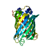
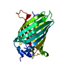
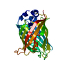
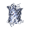
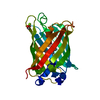
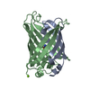
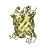
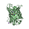
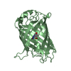
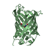
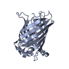
| Title | Cryogenic temperature structure of the fluorescent protein Cerulean recorded after an accumulated dose of 5.8 MGy | ||||||
 Components Components | Green fluorescent protein | ||||||
 Keywords Keywords | FLUORESCENT PROTEIN / tryptophan-based chromophore / Cerulean / hydrogen atoms | ||||||
| Function / homology | Green fluorescent protein, GFP / Green fluorescent protein-related / Green fluorescent protein / Green fluorescent protein / bioluminescence / generation of precursor metabolites and energy / CARBON DIOXIDE / Green fluorescent protein Function and homology information Function and homology information | ||||||
| Biological species |   Aequorea victoria (jellyfish) Aequorea victoria (jellyfish) | ||||||
| Method | X-RAY DIFFRACTION / SYNCHROTRON / Resolution: 1.82 Å | ||||||
 Authors Authors | Gotthard, G. / Aumonier, S. / Royant, A. | ||||||
 Citation Citation | Journal: Iucrj / Year: 2019 Title: Specific radiation damage is a lesser concern at room temperature. Authors: Gotthard, G. / Aumonier, S. / De Sanctis, D. / Leonard, G. / von Stetten, D. / Royant, A. | ||||||
| History |
|
- Structure visualization
| Structure viewer | Molecule: MolmilJmol/JSmol |
|---|
- Downloads & links
-Download
| PDBx/mmCIF format | 6qq9.cif.gz | 62.6 KB | Display | PDBx/mmCIF format |
|---|---|---|---|---|
| PDB format | pdb6qq9.ent.gz | 48.8 KB | Display | PDB format |
| PDBx/mmJSON format | 6qq9.json.gz | Tree view | PDBx/mmJSON format | |
| Others |  Other downloads Other downloads |
-Validation report
| Arichive directory | https://data.pdbj.org/pub/pdb/validation_reports/qq/6qq9ftp://data.pdbj.org/pub/pdb/validation_reports/qq/6qq9 | HTTPS FTP |
|---|
-Related structure data
| Related structure data | 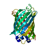 6qq8C 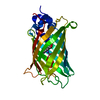 6qqaC 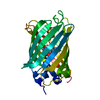 6qqbC 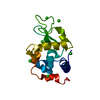 6qqcC 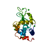 6qqdC  6qqeC 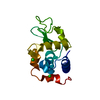 6qqfC 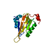 6qqhC 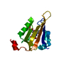 6qqiC 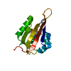 6qqjC 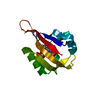 6qqkC 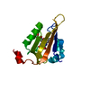 6qsaC C: citing same article ( |
|---|---|
| Similar structure data |


-Links
 PDBj
PDBj


- Assembly
| Deposited unit | 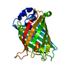
| ||||||||
|---|---|---|---|---|---|---|---|---|---|
| 1 |
| ||||||||
| Unit cell |
|
-Components
| #1: Protein | Mass: 26789.236 Da / Num. of mol.: 1 Source method: isolated from a genetically manipulated source Source: (gene. exp.) Aequorea victoria (jellyfish) / Gene: GFP / Production host:  Escherichia coli (E. coli) / References: UniProt: P42212 Escherichia coli (E. coli) / References: UniProt: P42212 |
|---|---|
| #2: Chemical | ChemComp-CO2 / Carbon dioxide  Mass: 44.010 Da / Num. of mol.: 1 / Source method: obtained synthetically / Formula: CO2 Mass: 44.010 Da / Num. of mol.: 1 / Source method: obtained synthetically / Formula: CO2 |
| #3: Water | ChemComp-HOH / Water Mass: 18.015 Da / Num. of mol.: 228 / Source method: isolated from a natural source / Formula: H2O Mass: 18.015 Da / Num. of mol.: 228 / Source method: isolated from a natural source / Formula: H2O |
-Experimental details
-Experiment
| Experiment | Method: X-RAY DIFFRACTION / Number of used crystals: 1 |
|---|
- Sample preparation
| Crystal | Density Matthews: 2.13 Å3/Da / Density % sol: 42.3 % |
|---|---|
| Crystal grow | Temperature: 293 K / Method: vapor diffusion, hanging drop / pH: 7 / Details: 100mM HEPES 7.0, 12% PEG8000, 100mM MgCl2 |
-Data collection
| Diffraction | Mean temperature: 100 K / Serial crystal experiment: N |
|---|---|
| Diffraction source | Source: SYNCHROTRON / Site: ESRF  / Beamline: ID29 / Wavelength: 0.976 Å / Beamline: ID29 / Wavelength: 0.976 Å |
| Detector | Type: DECTRIS PILATUS3 6M / Detector: PIXEL / Date: Dec 1, 2014 |
| Radiation | Monochromator: Si(311) / Protocol: SINGLE WAVELENGTH / Monochromatic (M) / Laue (L): M / Scattering type: x-ray |
| Radiation wavelength | Wavelength: 0.976 Å / Relative weight: 1 |
| Reflection | Resolution: 1.82→46.99 Å / Num. obs: 21133 / % possible obs: 99.9 % / Redundancy: 6.8 % / Biso Wilson estimate: 38.708 Å2 / CC1/2: 1 / Net I/σ(I): 28.23 |
| Reflection shell | Resolution: 1.82→1.87 Å / Redundancy: 7.3 % / Mean I/σ(I) obs: 2.05 / Num. unique obs: 1544 / CC1/2: 0.726 / % possible all: 100 |
- Processing
| Software |
| ||||||||||||||||||||||||||||||||||||||||||||||||||||||||||||||||||||||||||||||||||||||||||||||||||||||||||||||||||||||||||||||||||||||||||||||||||||||||||||||||||||||||||||||||||||||
|---|---|---|---|---|---|---|---|---|---|---|---|---|---|---|---|---|---|---|---|---|---|---|---|---|---|---|---|---|---|---|---|---|---|---|---|---|---|---|---|---|---|---|---|---|---|---|---|---|---|---|---|---|---|---|---|---|---|---|---|---|---|---|---|---|---|---|---|---|---|---|---|---|---|---|---|---|---|---|---|---|---|---|---|---|---|---|---|---|---|---|---|---|---|---|---|---|---|---|---|---|---|---|---|---|---|---|---|---|---|---|---|---|---|---|---|---|---|---|---|---|---|---|---|---|---|---|---|---|---|---|---|---|---|---|---|---|---|---|---|---|---|---|---|---|---|---|---|---|---|---|---|---|---|---|---|---|---|---|---|---|---|---|---|---|---|---|---|---|---|---|---|---|---|---|---|---|---|---|---|---|---|---|---|
| Refinement | Resolution: 1.82→46.99 Å / Cor.coef. Fo:Fc: 0.978 / Cor.coef. Fo:Fc free: 0.966 / SU B: 3.202 / SU ML: 0.093 / Cross valid method: THROUGHOUT / ESU R: 0.121 / ESU R Free: 0.118 / Details: HYDROGENS HAVE BEEN ADDED IN THE RIDING POSITIONS
| ||||||||||||||||||||||||||||||||||||||||||||||||||||||||||||||||||||||||||||||||||||||||||||||||||||||||||||||||||||||||||||||||||||||||||||||||||||||||||||||||||||||||||||||||||||||
| Solvent computation | Ion probe radii: 0.8 Å / Shrinkage radii: 0.8 Å / VDW probe radii: 1.2 Å | ||||||||||||||||||||||||||||||||||||||||||||||||||||||||||||||||||||||||||||||||||||||||||||||||||||||||||||||||||||||||||||||||||||||||||||||||||||||||||||||||||||||||||||||||||||||
| Displacement parameters | Biso mean: 33.889 Å2
| ||||||||||||||||||||||||||||||||||||||||||||||||||||||||||||||||||||||||||||||||||||||||||||||||||||||||||||||||||||||||||||||||||||||||||||||||||||||||||||||||||||||||||||||||||||||
| Refinement step | Cycle: 1 / Resolution: 1.82→46.99 Å
| ||||||||||||||||||||||||||||||||||||||||||||||||||||||||||||||||||||||||||||||||||||||||||||||||||||||||||||||||||||||||||||||||||||||||||||||||||||||||||||||||||||||||||||||||||||||
| Refine LS restraints |
|