 Movie
Movie Controller

[English] 日本語
 Yorodumi
- PDB-6qlg: Crystal structure of AnUbiX (PadA1) in complex with FMN and dimet... -
+ Open data

- Basic information
| Entry | Database: PDB / ID: 6qlg | |||||||||
|---|---|---|---|---|---|---|---|---|---|---|
| Title | Crystal structure of AnUbiX (PadA1) in complex with FMN and dimethylallyl pyrophosphate | |||||||||
 Components Components | Flavin prenyltransferase PAD1, mitochondrial | |||||||||
 Keywords Keywords |  TRANSFERASE / UbiX Prenyltransferase Flavin binding TRANSFERASE / UbiX Prenyltransferase Flavin binding | |||||||||
| Function / homology |  Function and homology informationflavin prenyltransferase / flavin prenyltransferase activity / mitochondrion Function and homology informationflavin prenyltransferase / flavin prenyltransferase activity / mitochondrionSimilarity search - Function | |||||||||
| Biological species |  Aspergillus niger (mold) Aspergillus niger (mold) | |||||||||
| Method | X-RAY DIFFRACTION / SYNCHROTRON / MOLECULAR REPLACEMENT / Resolution: 2.15 Å | |||||||||
 Authors Authors | Marshall, S.A. / Payne, K.A.P. / Leys, D. | |||||||||
| Funding support |  United Kingdom, 2items United Kingdom, 2items
| |||||||||
 Citation Citation | Journal: Nat Commun / Year: 2019 Title: The UbiX flavin prenyltransferase reaction mechanism resembles class I terpene cyclase chemistry. Authors: Marshall, S.A. / Payne, K.A.P. / Fisher, K. / White, M.D. / Ni Cheallaigh, A. / Balaikaite, A. / Rigby, S.E.J. / Leys, D. | |||||||||
| History |
|
- Structure visualization
| Structure viewer | Molecule: MolmilJmol/JSmol |
|---|
- Downloads & links
-Download
| PDBx/mmCIF format | 6qlg.cif.gz | 309.8 KB | Display | PDBx/mmCIF format |
|---|---|---|---|---|
| PDB format | pdb6qlg.ent.gz | 201.9 KB | Display | PDB format |
| PDBx/mmJSON format | 6qlg.json.gz | Tree view | PDBx/mmJSON format | |
| Others |  Other downloads Other downloads |
-Validation report
| Arichive directory | https://data.pdbj.org/pub/pdb/validation_reports/ql/6qlgftp://data.pdbj.org/pub/pdb/validation_reports/ql/6qlg | HTTPS FTP |
|---|
-Related structure data
| Related structure data |  6qlhC 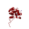 6qliC 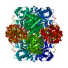 6qljC  6qlkC  6qllC  6qlvC C: citing same article ( |
|---|---|
| Similar structure data |


-Links
 PDBj
PDBj- Assembly
| Deposited unit | 
| ||||||||||||
|---|---|---|---|---|---|---|---|---|---|---|---|---|---|
| 1 | 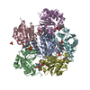
| ||||||||||||
| Unit cell |
| ||||||||||||
| Components on special symmetry positions |
|
-Components
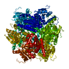
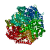
-Protein , 1 types, 6 molecules CABDEF
| #1: Protein | Mass: 25078.951 Da / Num. of mol.: 6 Source method: isolated from a genetically manipulated source Source: (gene. exp.) Aspergillus niger (mold) / Gene: PAD1, An03g06570 / Production host:  Escherichia coli BL21(DE3) (bacteria) / References: UniProt: A3F715, flavin prenyltransferase Escherichia coli BL21(DE3) (bacteria) / References: UniProt: A3F715, flavin prenyltransferase |
|---|
-Non-polymers , 5 types, 605 molecules 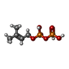


| #2: Chemical | ChemComp-DMA / Dimethylallyl pyrophosphate Mass: 246.092 Da / Num. of mol.: 6 / Source method: obtained synthetically / Formula: C5H12O7P2 Mass: 246.092 Da / Num. of mol.: 6 / Source method: obtained synthetically / Formula: C5H12O7P2#3: Chemical | ChemComp-FMN / Flavin mononucleotide Mass: 456.344 Da / Num. of mol.: 6 / Source method: obtained synthetically / Formula: C17H21N4O9P Mass: 456.344 Da / Num. of mol.: 6 / Source method: obtained synthetically / Formula: C17H21N4O9P#4: Chemical | ChemComp-PO4 / | Phosphate Mass: 94.971 Da / Num. of mol.: 1 / Source method: obtained synthetically / Formula: PO4 Mass: 94.971 Da / Num. of mol.: 1 / Source method: obtained synthetically / Formula: PO4#5: Chemical | Diethylene glycol Mass: 106.120 Da / Num. of mol.: 2 / Source method: obtained synthetically / Formula: C4H10O3 Mass: 106.120 Da / Num. of mol.: 2 / Source method: obtained synthetically / Formula: C4H10O3#6: Water | ChemComp-HOH / | WaterMass: 18.015 Da / Num. of mol.: 590 / Source method: isolated from a natural source / Formula: H2O |
|---|
-Experimental details
-Experiment
| Experiment | Method: X-RAY DIFFRACTION / Number of used crystals: 1 |
|---|
- Sample preparation
| Crystal | Density Matthews: 2.87 Å3/Da / Density % sol: 57.08 % |
|---|---|
| Crystal grow | Temperature: 294 K / Method: vapor diffusion, sitting drop Details: JCSG + D3 (Molecular Dimensions): 0.2 M NaCl, 0.1 M Na/K Phosphate pH 6.2, 30% v/v PEG200 |
-Data collection
| Diffraction | Mean temperature: 100 K / Serial crystal experiment: N |
|---|---|
| Diffraction source | Source: SYNCHROTRON / Site: Diamond / Beamline: I03 / Wavelength: 0.9763 Å |
| Detector | Type: DECTRIS PILATUS3 6M / Detector: PIXEL / Date: Jul 12, 2014 |
| Radiation | Protocol: SINGLE WAVELENGTH / Monochromatic (M) / Laue (L): M / Scattering type: x-ray |
| Radiation wavelength | Wavelength: 0.9763 Å / Relative weight: 1 |
| Reflection | Resolution: 2.15→95.7 Å / Num. obs: 80295 / % possible obs: 99.98 % / Redundancy: 14.7 % / Biso Wilson estimate: 29.17 Å2 / CC1/2: 0.998 / Rmerge(I) obs: 0.1247 / Net I/σ(I): 15.5 |
| Reflection shell | Resolution: 2.15→2.27 Å |
- Processing
| Software |
| ||||||||||||||||||||||||||||||||||||||||||||||||||||||||||||||||||||||||||||||||||||||||||||||||||||||||||||||||||||||||||||||||||||||||||||||||||||||||||||||||||||||||||||||||||||||||||||||||||||||||||||||||||
|---|---|---|---|---|---|---|---|---|---|---|---|---|---|---|---|---|---|---|---|---|---|---|---|---|---|---|---|---|---|---|---|---|---|---|---|---|---|---|---|---|---|---|---|---|---|---|---|---|---|---|---|---|---|---|---|---|---|---|---|---|---|---|---|---|---|---|---|---|---|---|---|---|---|---|---|---|---|---|---|---|---|---|---|---|---|---|---|---|---|---|---|---|---|---|---|---|---|---|---|---|---|---|---|---|---|---|---|---|---|---|---|---|---|---|---|---|---|---|---|---|---|---|---|---|---|---|---|---|---|---|---|---|---|---|---|---|---|---|---|---|---|---|---|---|---|---|---|---|---|---|---|---|---|---|---|---|---|---|---|---|---|---|---|---|---|---|---|---|---|---|---|---|---|---|---|---|---|---|---|---|---|---|---|---|---|---|---|---|---|---|---|---|---|---|---|---|---|---|---|---|---|---|---|---|---|---|---|---|---|---|---|
| Refinement | Method to determine structure: MOLECULAR REPLACEMENT / Resolution: 2.15→95.7 Å / SU ML: 0.1838 / Cross valid method: FREE R-VALUE / σ(F): 1.34 / Phase error: 17.881
| ||||||||||||||||||||||||||||||||||||||||||||||||||||||||||||||||||||||||||||||||||||||||||||||||||||||||||||||||||||||||||||||||||||||||||||||||||||||||||||||||||||||||||||||||||||||||||||||||||||||||||||||||||
| Solvent computation | Shrinkage radii: 0.9 Å / VDW probe radii: 1.11 Å | ||||||||||||||||||||||||||||||||||||||||||||||||||||||||||||||||||||||||||||||||||||||||||||||||||||||||||||||||||||||||||||||||||||||||||||||||||||||||||||||||||||||||||||||||||||||||||||||||||||||||||||||||||
| Displacement parameters | Biso mean: 33.66 Å2 | ||||||||||||||||||||||||||||||||||||||||||||||||||||||||||||||||||||||||||||||||||||||||||||||||||||||||||||||||||||||||||||||||||||||||||||||||||||||||||||||||||||||||||||||||||||||||||||||||||||||||||||||||||
| Refinement step | Cycle: LAST / Resolution: 2.15→95.7 Å
| ||||||||||||||||||||||||||||||||||||||||||||||||||||||||||||||||||||||||||||||||||||||||||||||||||||||||||||||||||||||||||||||||||||||||||||||||||||||||||||||||||||||||||||||||||||||||||||||||||||||||||||||||||
| Refine LS restraints |
| ||||||||||||||||||||||||||||||||||||||||||||||||||||||||||||||||||||||||||||||||||||||||||||||||||||||||||||||||||||||||||||||||||||||||||||||||||||||||||||||||||||||||||||||||||||||||||||||||||||||||||||||||||
| LS refinement shell |
|
