 Movie
Movie Controller


+ Open data

- Basic information
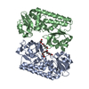
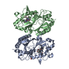
| Entry | Database: PDB / ID: 6qau | ||||||
|---|---|---|---|---|---|---|---|
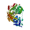
| Title | Crystal structure of ULK2 in complexed with MRT67307 | ||||||
 Components Components | Serine/threonine-protein kinase ULK2 | ||||||
 Keywords Keywords |  TRANSFERASE / ULK2 / autophagy / kinase / inhibitor complex / Structural Genomics / Structural Genomics Consortium / SGC TRANSFERASE / ULK2 / autophagy / kinase / inhibitor complex / Structural Genomics / Structural Genomics Consortium / SGC | ||||||
| Function / homology |  Function and homology information Function and homology informationnegative regulation of collateral sprouting / collateral sprouting / phagophore assembly site membrane / piecemeal microautophagy of the nucleus / phagophore assembly site / axon extension / reticulophagy / response to starvation / autophagosome assembly / autophagosome ...negative regulation of collateral sprouting / collateral sprouting / phagophore assembly site membrane / piecemeal microautophagy of the nucleus / phagophore assembly site / axon extension / reticulophagy / response to starvation / autophagosome assembly / autophagosome / positive regulation of autophagy / cytoplasmic vesicle membrane / autophagy / protein autophosphorylation / non-specific serine/threonine protein kinase / protein serine kinase activity / protein serine/threonine kinase activity / signal transduction / ATP binding / cytosol / cytoplasmSimilarity search - Function | ||||||
| Biological species |  Homo sapiens (human) Homo sapiens (human) | ||||||
| Method | X-RAY DIFFRACTION / SYNCHROTRON / MOLECULAR REPLACEMENT / Resolution: 2.48 Å | ||||||
 Authors Authors | Chaikuad, A. / Arrowsmith, C.H. / Edwards, A.M. / Bountra, C. / Knapp, S. / Structural Genomics Consortium / Structural Genomics Consortium (SGC) | ||||||
 Citation Citation | Journal: Biochem.J. / Year: 2019 Title: Conservation of structure, function and inhibitor binding in UNC-51-like kinase 1 and 2 (ULK1/2). Authors: Chaikuad, A. / Koschade, S.E. / Stolz, A. / Zivkovic, K. / Pohl, C. / Shaid, S. / Ren, H. / Lambert, L.J. / Cosford, N.D.P. / Brandts, C.H. / Knapp, S. | ||||||
| History |
|
- Structure visualization
| Structure viewer | Molecule: MolmilJmol/JSmol |
|---|
- Downloads & links
-Download
| PDBx/mmCIF format | 6qau.cif.gz | 335.5 KB | Display | PDBx/mmCIF format |
|---|---|---|---|---|
| PDB format | pdb6qau.ent.gz | 275.4 KB | Display | PDB format |
| PDBx/mmJSON format | 6qau.json.gz | Tree view | PDBx/mmJSON format | |
| Others |  Other downloads Other downloads |
-Validation report
| Arichive directory | https://data.pdbj.org/pub/pdb/validation_reports/qa/6qauftp://data.pdbj.org/pub/pdb/validation_reports/qa/6qau | HTTPS FTP |
|---|
-Related structure data
| Related structure data | 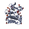 6qasC 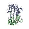 6qatC 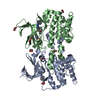 6qavC 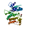 4wnoS S: Starting model for refinement C: citing same article ( |
|---|---|
| Similar structure data |


-Links
 PDBj
PDBj
- Assembly
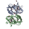
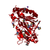
| Deposited unit | 
| |||||||||||||||||||||||||||||||||||||||||||||||||||||||||||||||||||||||||||||||||
|---|---|---|---|---|---|---|---|---|---|---|---|---|---|---|---|---|---|---|---|---|---|---|---|---|---|---|---|---|---|---|---|---|---|---|---|---|---|---|---|---|---|---|---|---|---|---|---|---|---|---|---|---|---|---|---|---|---|---|---|---|---|---|---|---|---|---|---|---|---|---|---|---|---|---|---|---|---|---|---|---|---|---|
| 1 | 
| |||||||||||||||||||||||||||||||||||||||||||||||||||||||||||||||||||||||||||||||||
| 2 | 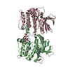
| |||||||||||||||||||||||||||||||||||||||||||||||||||||||||||||||||||||||||||||||||
| Unit cell |
| |||||||||||||||||||||||||||||||||||||||||||||||||||||||||||||||||||||||||||||||||
| Components on special symmetry positions |
| |||||||||||||||||||||||||||||||||||||||||||||||||||||||||||||||||||||||||||||||||
| Noncrystallographic symmetry (NCS) | NCS domain:
NCS domain segments: Component-ID: 0 / Beg auth comp-ID: SER / Beg label comp-ID: SER / Refine code: 0
NCS ensembles :
|
-Components
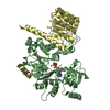
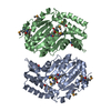
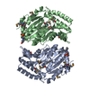
-Protein , 1 types, 3 molecules ABC
| #1: Protein | Mass: 31767.721 Da / Num. of mol.: 3 Source method: isolated from a genetically manipulated source Source: (gene. exp.) Homo sapiens (human) / Gene: ULK2, KIAA0623 / Plasmid: pNIC28-Bsa4 / Production host:  Escherichia coli BL21(DE3) (bacteria) / Variant (production host): R3-pRARE2 Escherichia coli BL21(DE3) (bacteria) / Variant (production host): R3-pRARE2References: UniProt: Q8IYT8, non-specific serine/threonine protein kinase |
|---|
-Non-polymers , 5 types, 188 molecules 


| #2: Chemical |  Mass: 464.603 Da / Num. of mol.: 3 / Source method: obtained synthetically / Formula: C26H36N6O2 Mass: 464.603 Da / Num. of mol.: 3 / Source method: obtained synthetically / Formula: C26H36N6O2#3: Chemical | ChemComp-EDO / Ethylene glycol Mass: 62.068 Da / Num. of mol.: 9 / Source method: obtained synthetically / Formula: C2H6O2 Mass: 62.068 Da / Num. of mol.: 9 / Source method: obtained synthetically / Formula: C2H6O2#4: Chemical | ChemComp-GOL / | Glycerol Mass: 92.094 Da / Num. of mol.: 1 / Source method: obtained synthetically / Formula: C3H8O3 Mass: 92.094 Da / Num. of mol.: 1 / Source method: obtained synthetically / Formula: C3H8O3#5: Chemical | ChemComp-CL / | Chloride Mass: 35.453 Da / Num. of mol.: 1 / Source method: obtained synthetically / Formula: Cl Mass: 35.453 Da / Num. of mol.: 1 / Source method: obtained synthetically / Formula: Cl#6: Water | ChemComp-HOH / | WaterMass: 18.015 Da / Num. of mol.: 174 / Source method: isolated from a natural source / Formula: H2O |
|---|
-Experimental details
-Experiment
| Experiment | Method: X-RAY DIFFRACTION / Number of used crystals: 1 |
|---|
- Sample preparation
| Crystal | Density Matthews: 2.93 Å3/Da / Density % sol: 58.07 % |
|---|---|
| Crystal grow | Temperature: 293.15 K / Method: vapor diffusion, sitting drop Details: 22.5% PEG 3350, 0.2 M sodium citrate, pH 5.9, 0.1 M bis-tris, pH 5.75, 5% glycerol PH range: 5.75-5.9 |
-Data collection
| Diffraction | Mean temperature: 100 K / Serial crystal experiment: N |
|---|---|
| Diffraction source | Source: SYNCHROTRON / Site: SLS  / Beamline: X06DA / Wavelength: 1 Å / Beamline: X06DA / Wavelength: 1 Å |
| Detector | Type: DECTRIS PILATUS3 2M / Detector: PIXEL / Date: Jun 8, 2018 |
| Radiation | Protocol: SINGLE WAVELENGTH / Monochromatic (M) / Laue (L): M / Scattering type: x-ray |
| Radiation wavelength | Wavelength: 1 Å / Relative weight: 1 |
| Reflection | Resolution: 2.48→58.5 Å / Num. obs: 40240 / % possible obs: 100 % / Redundancy: 9.8 % / Biso Wilson estimate: 47 Å2 / CC1/2: 0.998 / Rmerge(I) obs: 0.136 / Net I/σ(I): 11.5 |
| Reflection shell | Resolution: 2.48→2.61 Å / Redundancy: 9.8 % / Rmerge(I) obs: 0.998 / Mean I/σ(I) obs: 2.2 / Num. unique obs: 5786 / CC1/2: 0.711 / % possible all: 100 |
- Processing
| Software |
| ||||||||||||||||||||||||||||||||||||||||||||||||||||||||||||||||||||||||||||||||||||||||||||||||||||||||||||||||||||||||||||||||||||||||||||||||||||||||||||||||||||||||||||||||||||||
|---|---|---|---|---|---|---|---|---|---|---|---|---|---|---|---|---|---|---|---|---|---|---|---|---|---|---|---|---|---|---|---|---|---|---|---|---|---|---|---|---|---|---|---|---|---|---|---|---|---|---|---|---|---|---|---|---|---|---|---|---|---|---|---|---|---|---|---|---|---|---|---|---|---|---|---|---|---|---|---|---|---|---|---|---|---|---|---|---|---|---|---|---|---|---|---|---|---|---|---|---|---|---|---|---|---|---|---|---|---|---|---|---|---|---|---|---|---|---|---|---|---|---|---|---|---|---|---|---|---|---|---|---|---|---|---|---|---|---|---|---|---|---|---|---|---|---|---|---|---|---|---|---|---|---|---|---|---|---|---|---|---|---|---|---|---|---|---|---|---|---|---|---|---|---|---|---|---|---|---|---|---|---|---|
| Refinement | Method to determine structure: MOLECULAR REPLACEMENT Starting model: 4WNO Resolution: 2.48→58.5 Å / Cor.coef. Fo:Fc: 0.948 / Cor.coef. Fo:Fc free: 0.911 / SU B: 19.941 / SU ML: 0.217 / Cross valid method: THROUGHOUT / ESU R: 0.376 / ESU R Free: 0.25 / Details: HYDROGENS HAVE BEEN ADDED IN THE RIDING POSITIONS
| ||||||||||||||||||||||||||||||||||||||||||||||||||||||||||||||||||||||||||||||||||||||||||||||||||||||||||||||||||||||||||||||||||||||||||||||||||||||||||||||||||||||||||||||||||||||
| Solvent computation | Ion probe radii: 0.8 Å / Shrinkage radii: 0.8 Å / VDW probe radii: 1.2 Å | ||||||||||||||||||||||||||||||||||||||||||||||||||||||||||||||||||||||||||||||||||||||||||||||||||||||||||||||||||||||||||||||||||||||||||||||||||||||||||||||||||||||||||||||||||||||
| Displacement parameters | Biso mean: 57.774 Å2
| ||||||||||||||||||||||||||||||||||||||||||||||||||||||||||||||||||||||||||||||||||||||||||||||||||||||||||||||||||||||||||||||||||||||||||||||||||||||||||||||||||||||||||||||||||||||
| Refine analyze | Luzzati coordinate error obs: 0.346 Å | ||||||||||||||||||||||||||||||||||||||||||||||||||||||||||||||||||||||||||||||||||||||||||||||||||||||||||||||||||||||||||||||||||||||||||||||||||||||||||||||||||||||||||||||||||||||
| Refinement step | Cycle: 1 / Resolution: 2.48→58.5 Å
| ||||||||||||||||||||||||||||||||||||||||||||||||||||||||||||||||||||||||||||||||||||||||||||||||||||||||||||||||||||||||||||||||||||||||||||||||||||||||||||||||||||||||||||||||||||||
| Refine LS restraints |
|