- PDB-6all: Crystal structure of a predicted ferric/iron (III) hydroxymate si... -
+
Open data
ID or keywords:
Loading...
-
Basic information
Entry
Database: PDB / ID: 6all
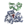
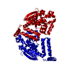
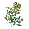
Title
Crystal structure of a predicted ferric/iron (III) hydroxymate siderophore substrate binding protein from Bacillus anthracis
Components
Fe(3+)-citrate-binding protein yfmC
Keywords
METAL TRANSPORT / structural genomics / NIAID / national institute of allergy and infectious diseases / PERIPLASMIC LIGAND BINDING PROTEIN / SUBSTRATE BINDING PROTEIN / ABC TRANSPORT PROTEIN / alpha/beta protein / Center for Structural Genomics of Infectious Diseases / CSGID
Function / homology
Function and homology information
: / ATPase-coupled transmembrane transporter activity / iron ion transport / iron ion binding / plasma membrane Similarity search - Function
ABC transporter periplasmic binding domain / Periplasmic binding protein / Iron siderophore/cobalamin periplasmic-binding domain profile. / Nitrogenase molybdenum iron protein domain / Prokaryotic membrane lipoprotein lipid attachment site profile. / Rossmann fold / 3-Layer(aba) Sandwich / Alpha Beta Similarity search - Domain/homology
National Institutes of Health/National Institute Of Allergy and Infectious Diseases (NIH/NIAID)
HHSN272201200026C
United States
Citation
Journal: To Be Published Title: Crystal structure of a predicted ferric/iron (III) hydroxymate siderophore substrate binding protein from Bacillus anthracis Authors: Stogios, P.J.
In the structure databanks used in Yorodumi, some data are registered as the other names, "COVID-19 virus" and "2019-nCoV". Here are the details of the virus and the list of structure data.
Jan 31, 2019. EMDB accession codes are about to change! (news from PDBe EMDB page)
EMDB accession codes are about to change! (news from PDBe EMDB page)
The allocation of 4 digits for EMDB accession codes will soon come to an end. Whilst these codes will remain in use, new EMDB accession codes will include an additional digit and will expand incrementally as the available range of codes is exhausted. The current 4-digit format prefixed with “EMD-” (i.e. EMD-XXXX) will advance to a 5-digit format (i.e. EMD-XXXXX), and so on. It is currently estimated that the 4-digit codes will be depleted around Spring 2019, at which point the 5-digit format will come into force.
The EM Navigator/Yorodumi systems omit the EMD- prefix.
Related info.:Q: What is EMD? / ID/Accession-code notation in Yorodumi/EM Navigator
Yorodumi is a browser for structure data from EMDB, PDB, SASBDB, etc.
This page is also the successor to EM Navigator detail page, and also detail information page/front-end page for Omokage search.
The word "yorodu" (or yorozu) is an old Japanese word meaning "ten thousand". "mi" (miru) is to see.
Related info.:EMDB / PDB / SASBDB / Comparison of 3 databanks / Yorodumi Search / Aug 31, 2016. New EM Navigator & Yorodumi / Yorodumi Papers / Jmol/JSmol / Function and homology information / Changes in new EM Navigator and Yorodumi
 Movie
Movie Controller

 Yorodumi
 Open data
 Basic information
 Components
 Keywords
 structural genomics / NIAID /
  Function and homology information
 Authors
 United States, 1items
  Citation
 Structure visualization
 Downloads & links
 Other downloads
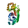


 PDBj
PDBj


 Assembly
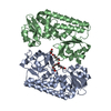
 Mass: 634.751 Da / Num. of mol.: 3 / Source method: obtained synthetically / Formula: C28H58O15
 Mass: 18.015 Da / Num. of mol.: 49 / Source method: isolated from a natural source / Formula: H2O
 Sample preparation
 Processing