 Movie
Movie Controller

[English] 日本語
 Yorodumi
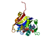
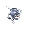
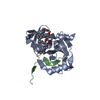
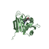
- PDB-6pra: S. aureus dihydrofolate reductase with NADP(H) and empty folate pocket -
+ Open data

- Basic information
| Entry | Database: PDB / ID: 6pra | ||||||
|---|---|---|---|---|---|---|---|
| Title | S. aureus dihydrofolate reductase with NADP(H) and empty folate pocket | ||||||
 Components Components | Dihydrofolate reductase | ||||||
 Keywords Keywords | OXIDOREDUCTASE/INHIBITOR / DIHYDROFOLATE REDUCTASE / OXIDOREDUCTASE-INHIBITOR COMPLEX | ||||||
| Function / homology |  Function and homology information Function and homology informationdihydrofolate metabolic process / glycine biosynthetic process / dihydrofolate reductase / folic acid metabolic process / dihydrofolate reductase activity / tetrahydrofolate biosynthetic process / one-carbon metabolic process / NADP binding / cytosolSimilarity search - Function | ||||||
| Biological species |   Staphylococcus aureus (bacteria) Staphylococcus aureus (bacteria) | ||||||
| Method | X-RAY DIFFRACTION / MOLECULAR REPLACEMENT / Resolution: 2.01 Å | ||||||
 Authors Authors | Bourne, C.R. / Thomas, L.M. | ||||||
| Funding support |  United States, 1items United States, 1items
| ||||||
 Citation Citation | Journal: Eur.J.Med.Chem. / Year: 2020 Title: Inhibitor design to target a unique feature in the folate pocket of Staphylococcus aureus dihydrofolate reductase. Authors: Muddala, N.P. / White, J.C. / Nammalwar, B. / Pratt, I. / Thomas, L.M. / Bunce, R.A. / Berlin, K.D. / Bourne, C.R. | ||||||
| History |
|
- Structure visualization
| Structure viewer | Molecule: MolmilJmol/JSmol |
|---|
- Downloads & links
-Download
| PDBx/mmCIF format | 6pra.cif.gz | 55.8 KB | Display | PDBx/mmCIF format |
|---|---|---|---|---|
| PDB format | pdb6pra.ent.gz | 37.1 KB | Display | PDB format |
| PDBx/mmJSON format | 6pra.json.gz | Tree view | PDBx/mmJSON format | |
| Others |  Other downloads Other downloads |
-Validation report
| Arichive directory | https://data.pdbj.org/pub/pdb/validation_reports/pr/6praftp://data.pdbj.org/pub/pdb/validation_reports/pr/6pra | HTTPS FTP |
|---|
-Related structure data
| Related structure data | 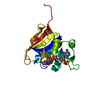 6pr6C  6pr7C  6pr8C 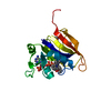 6pr9C 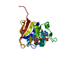 6prbC 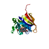 6prdC  3m08S S: Starting model for refinement C: citing same article ( |
|---|---|
| Similar structure data |


-Links
 PDBj
PDBj
- Assembly
| Deposited unit | 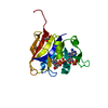
| ||||||||||
|---|---|---|---|---|---|---|---|---|---|---|---|
| 1 |
| ||||||||||
| Unit cell |
|
-Components
| #1: Protein | / DHFR Mass: 18742.533 Da / Num. of mol.: 1 Source method: isolated from a genetically manipulated source Source: (gene. exp.) Staphylococcus aureus (bacteria) / Gene: folA / Production host: Escherichia coli (E. coli) / References: UniProt: P0A017, dihydrofolate reductase | ||||
|---|---|---|---|---|---|
| #2: Chemical | ChemComp-NAP / Nicotinamide adenine dinucleotide phosphate  Mass: 743.405 Da / Num. of mol.: 1 / Source method: obtained synthetically / Formula: C21H28N7O17P3 Mass: 743.405 Da / Num. of mol.: 1 / Source method: obtained synthetically / Formula: C21H28N7O17P3 | ||||
| #3: Chemical | Glycerol  Mass: 92.094 Da / Num. of mol.: 2 / Source method: isolated from a natural source / Formula: C3H8O3 Mass: 92.094 Da / Num. of mol.: 2 / Source method: isolated from a natural source / Formula: C3H8O3#4: Water | ChemComp-HOH / | Water Mass: 18.015 Da / Num. of mol.: 149 / Source method: isolated from a natural source / Formula: H2O Mass: 18.015 Da / Num. of mol.: 149 / Source method: isolated from a natural source / Formula: H2OHas ligand of interest | N | |
-Experimental details
-Experiment
| Experiment | Method: X-RAY DIFFRACTION / Number of used crystals: 1 |
|---|
- Sample preparation
| Crystal | Density Matthews: 2.58 Å3/Da / Density % sol: 52.31 % |
|---|---|
| Crystal grow | Temperature: 293 K / Method: vapor diffusion / Details: 20% PEG6000, 0.1M MES, 0.15M Na Acetate |
-Data collection
| Diffraction | Mean temperature: 100 K / Serial crystal experiment: N |
|---|---|
| Diffraction source | Source: ROTATING ANODE / Type: RIGAKU RUH3R / Wavelength: 1.5418 Å |
| Detector | Type: RIGAKU RAXIS IV++ / Detector: IMAGE PLATE / Date: Apr 17, 2012 / Details: Osmic mirrors |
| Radiation | Monochromator: Mirrors / Protocol: SINGLE WAVELENGTH / Monochromatic (M) / Laue (L): M / Scattering type: x-ray |
| Radiation wavelength | Wavelength: 1.5418 Å / Relative weight: 1 |
| Reflection | Resolution: 2.01→25.101 Å / Num. obs: 13688 / % possible obs: 99.8 % / Redundancy: 11.2 % / Biso Wilson estimate: 20.37 Å2 / Rmerge(I) obs: 0.085 / Net I/σ(I): 13.5 |
| Reflection shell | Resolution: 2.01→2.08 Å / Rmerge(I) obs: 0.36 / Num. unique obs: 1331 |
- Processing
| Software |
| ||||||||||||||||||||||||||||||
|---|---|---|---|---|---|---|---|---|---|---|---|---|---|---|---|---|---|---|---|---|---|---|---|---|---|---|---|---|---|---|---|
| Refinement | Method to determine structure: MOLECULAR REPLACEMENT Starting model: 3M08 Resolution: 2.01→25.101 Å / SU ML: 0.19 / Cross valid method: THROUGHOUT / σ(F): 1.34 / Phase error: 20.16
| ||||||||||||||||||||||||||||||
| Solvent computation | Shrinkage radii: 0.9 Å / VDW probe radii: 1.11 Å | ||||||||||||||||||||||||||||||
| Displacement parameters | Biso max: 169.17 Å2 / Biso mean: 23.68 Å2 / Biso min: 6.46 Å2 | ||||||||||||||||||||||||||||||
| Refinement step | Cycle: final / Resolution: 2.01→25.101 Å
| ||||||||||||||||||||||||||||||
| Refine LS restraints |
| ||||||||||||||||||||||||||||||
| LS refinement shell | Refine-ID: X-RAY DIFFRACTION / Rfactor Rfree error: 0 / % reflection obs: 100 %
|
