 Movie
Movie Controller

[English] 日本語
 Yorodumi
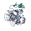
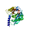
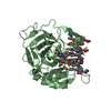
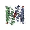
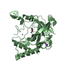
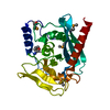
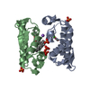
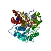
- PDB-6oze: Crystal structure of the catalytic domain of human Endonuclease V... -
+ Open data

- Basic information
| Entry | Database: PDB / ID: 6oze | ||||||
|---|---|---|---|---|---|---|---|
| Title | Crystal structure of the catalytic domain of human Endonuclease V (C140S/C225S/C226A/C228S) | ||||||
 Components Components | Endonuclease V | ||||||
 Keywords Keywords | HYDROLASE / Nucleic acid hydrolysis / RNA recognition / metal ion dependent catalysis / DNA damage / adenosine deamination | ||||||
| Function / homology |  Function and homology information Function and homology informationRNA endonuclease activity, producing 5'-phosphomonoesters / endodeoxyribonuclease activity, producing 5'-phosphomonoesters / Hydrolases; Acting on ester bonds; Endoribonucleases producing 5'-phosphomonoesters / cytoplasmic stress granule / single-stranded RNA binding / DNA repair / nucleolus / magnesium ion binding / DNA binding / cytoplasmSimilarity search - Function | ||||||
| Biological species |  Homo sapiens (human) Homo sapiens (human) | ||||||
| Method | X-RAY DIFFRACTION / SYNCHROTRON / MOLECULAR REPLACEMENT / Resolution: 1.5 Å | ||||||
 Authors Authors | Samara, N.L. / Yang, W. | ||||||
 Citation Citation | Journal: Mol.Cell / Year: 2019 Title: Evolution of Inosine-Specific Endonuclease V from Bacterial DNase to Eukaryotic RNase. Authors: Wu, J. / Samara, N.L. / Kuraoka, I. / Yang, W. | ||||||
| History |
|
- Structure visualization
| Structure viewer | Molecule: MolmilJmol/JSmol |
|---|
- Downloads & links
-Download
| PDBx/mmCIF format | 6oze.cif.gz | 121.8 KB | Display | PDBx/mmCIF format |
|---|---|---|---|---|
| PDB format | pdb6oze.ent.gz | 92.4 KB | Display | PDB format |
| PDBx/mmJSON format | 6oze.json.gz | Tree view | PDBx/mmJSON format | |
| Others |  Other downloads Other downloads |
-Validation report
| Arichive directory | https://data.pdbj.org/pub/pdb/validation_reports/oz/6ozeftp://data.pdbj.org/pub/pdb/validation_reports/oz/6oze | HTTPS FTP |
|---|
-Related structure data
| Related structure data |  6ozfC 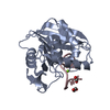 6ozgC 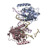 6ozhC 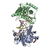 6oziC 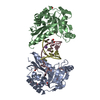 6ozjC 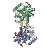 6ozkC 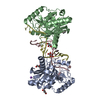 6ozlC 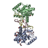 6ozmC 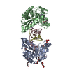 6oznC 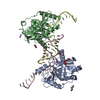 6ozoC  6ozpC 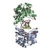 6ozqC  6ozrC 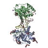 6ozsC  2w35S S: Starting model for refinement C: citing same article ( |
|---|---|
| Similar structure data |


-Links
 PDBj
PDBj- Assembly
| Deposited unit | 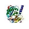
| ||||||||
|---|---|---|---|---|---|---|---|---|---|
| 1 |
| ||||||||
| Unit cell |
|
-Components
| #1: Protein | / hEndoV / Inosine-specific endoribonuclease Mass: 27157.359 Da / Num. of mol.: 1 / Mutation: C140S/C225S/C226A/C228S Source method: isolated from a genetically manipulated source Source: (gene. exp.) Homo sapiens (human) / Gene: ENDOV / Production host:  Escherichia coli (E. coli) Escherichia coli (E. coli)References: UniProt: Q8N8Q3, Hydrolases; Acting on ester bonds; Endoribonucleases producing 5'-phosphomonoesters | ||||||
|---|---|---|---|---|---|---|---|
| #2: Chemical | Acetate  Mass: 59.044 Da / Num. of mol.: 2 / Source method: obtained synthetically / Formula: C2H3O2 Mass: 59.044 Da / Num. of mol.: 2 / Source method: obtained synthetically / Formula: C2H3O2#3: Chemical | Glycerol  Mass: 92.094 Da / Num. of mol.: 2 / Source method: obtained synthetically / Formula: C3H8O3 Mass: 92.094 Da / Num. of mol.: 2 / Source method: obtained synthetically / Formula: C3H8O3#4: Chemical |   Mass: 22.990 Da / Num. of mol.: 2 / Source method: obtained synthetically / Formula: Na Mass: 22.990 Da / Num. of mol.: 2 / Source method: obtained synthetically / Formula: Na#5: Water | ChemComp-HOH / | Water Mass: 18.015 Da / Num. of mol.: 146 / Source method: isolated from a natural source / Formula: H2O Mass: 18.015 Da / Num. of mol.: 146 / Source method: isolated from a natural source / Formula: H2O |
-Experimental details
-Experiment
| Experiment | Method: X-RAY DIFFRACTION / Number of used crystals: 1 |
|---|
- Sample preparation
| Crystal | Density Matthews: 2.84 Å3/Da / Density % sol: 56.71 % |
|---|---|
| Crystal grow | Temperature: 293 K / Method: vapor diffusion, hanging drop / Details: 3.6 M sodium acetate |
-Data collection
| Diffraction | Mean temperature: 100 K / Serial crystal experiment: N | |||||||||||||||||||||
|---|---|---|---|---|---|---|---|---|---|---|---|---|---|---|---|---|---|---|---|---|---|---|
| Diffraction source | Source: SYNCHROTRON / Site: APS  / Beamline: 22-BM / Wavelength: 1 Å / Beamline: 22-BM / Wavelength: 1 Å | |||||||||||||||||||||
| Detector | Type: MARMOSAIC 225 mm CCD / Detector: CCD / Date: Mar 7, 2013 | |||||||||||||||||||||
| Radiation | Monochromator: double crystal Si(111) / Protocol: SINGLE WAVELENGTH / Monochromatic (M) / Laue (L): M / Scattering type: x-ray | |||||||||||||||||||||
| Radiation wavelength | Wavelength: 1 Å / Relative weight: 1 | |||||||||||||||||||||
| Reflection | Resolution: 1.5→24.73 Å / Num. obs: 47096 / % possible obs: 98.2 % / Redundancy: 1 % / Biso Wilson estimate: 21.1 Å2 / Net I/σ(I): 9.9 / Num. measured all: 47096 | |||||||||||||||||||||
| Reflection shell |
|
- Processing
| Software |
| ||||||||||||||||||||||||||||||||||||||||||||||||||||||||||||||||||||||||||||||||||||||||||||||||||||||||||||||||||||||||||||||||||||||||||||||||||||||||||||||||||||||||||||||||||||||||||||||||||||||||
|---|---|---|---|---|---|---|---|---|---|---|---|---|---|---|---|---|---|---|---|---|---|---|---|---|---|---|---|---|---|---|---|---|---|---|---|---|---|---|---|---|---|---|---|---|---|---|---|---|---|---|---|---|---|---|---|---|---|---|---|---|---|---|---|---|---|---|---|---|---|---|---|---|---|---|---|---|---|---|---|---|---|---|---|---|---|---|---|---|---|---|---|---|---|---|---|---|---|---|---|---|---|---|---|---|---|---|---|---|---|---|---|---|---|---|---|---|---|---|---|---|---|---|---|---|---|---|---|---|---|---|---|---|---|---|---|---|---|---|---|---|---|---|---|---|---|---|---|---|---|---|---|---|---|---|---|---|---|---|---|---|---|---|---|---|---|---|---|---|---|---|---|---|---|---|---|---|---|---|---|---|---|---|---|---|---|---|---|---|---|---|---|---|---|---|---|---|---|---|---|---|---|
| Refinement | Method to determine structure: MOLECULAR REPLACEMENT Starting model: PDB entry 2W35 Resolution: 1.5→24.729 Å / SU ML: 0.13 / Cross valid method: THROUGHOUT / σ(F): 1.97 / Phase error: 22.84
| ||||||||||||||||||||||||||||||||||||||||||||||||||||||||||||||||||||||||||||||||||||||||||||||||||||||||||||||||||||||||||||||||||||||||||||||||||||||||||||||||||||||||||||||||||||||||||||||||||||||||
| Solvent computation | Shrinkage radii: 0.9 Å / VDW probe radii: 1.11 Å | ||||||||||||||||||||||||||||||||||||||||||||||||||||||||||||||||||||||||||||||||||||||||||||||||||||||||||||||||||||||||||||||||||||||||||||||||||||||||||||||||||||||||||||||||||||||||||||||||||||||||
| Displacement parameters | Biso max: 110.49 Å2 / Biso mean: 37.4917 Å2 / Biso min: 14.73 Å2 | ||||||||||||||||||||||||||||||||||||||||||||||||||||||||||||||||||||||||||||||||||||||||||||||||||||||||||||||||||||||||||||||||||||||||||||||||||||||||||||||||||||||||||||||||||||||||||||||||||||||||
| Refinement step | Cycle: final / Resolution: 1.5→24.729 Å
| ||||||||||||||||||||||||||||||||||||||||||||||||||||||||||||||||||||||||||||||||||||||||||||||||||||||||||||||||||||||||||||||||||||||||||||||||||||||||||||||||||||||||||||||||||||||||||||||||||||||||
| Refine LS restraints |
| ||||||||||||||||||||||||||||||||||||||||||||||||||||||||||||||||||||||||||||||||||||||||||||||||||||||||||||||||||||||||||||||||||||||||||||||||||||||||||||||||||||||||||||||||||||||||||||||||||||||||
| LS refinement shell | Refine-ID: X-RAY DIFFRACTION / Rfactor Rfree error: 0 / Total num. of bins used: 16
| ||||||||||||||||||||||||||||||||||||||||||||||||||||||||||||||||||||||||||||||||||||||||||||||||||||||||||||||||||||||||||||||||||||||||||||||||||||||||||||||||||||||||||||||||||||||||||||||||||||||||
| Refinement TLS params. | Method: refined / Refine-ID: X-RAY DIFFRACTION
| ||||||||||||||||||||||||||||||||||||||||||||||||||||||||||||||||||||||||||||||||||||||||||||||||||||||||||||||||||||||||||||||||||||||||||||||||||||||||||||||||||||||||||||||||||||||||||||||||||||||||
| Refinement TLS group |
|
