 Movie
Movie Controller

[English] 日本語
 Yorodumi
- PDB-3rq2: Crystal Structure of ADP/ATP-dependent NAD(P)H-hydrate dehydratas... -
+ Open data

- Basic information
| Entry | Database: PDB / ID: 3rq2 | ||||||
|---|---|---|---|---|---|---|---|
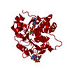
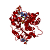
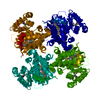
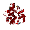
| Title | Crystal Structure of ADP/ATP-dependent NAD(P)H-hydrate dehydratase from Bacillus subtilis co-crystallized with ATP/Mg2+ and soaked with NADH | ||||||
 Components Components | ADP/ATP-dependent NAD(P)H-hydrate dehydratase | ||||||
 Keywords Keywords | lyase/lyase substrate /  STRUCTURAL GENOMICS / PSI-biology / PROTEIN STRUCTURE INITIATIVE / MIDWEST CENTER FOR STRUCTURAL GENOMICS / MCSG / LYASE / lyase-lyase substrate complex STRUCTURAL GENOMICS / PSI-biology / PROTEIN STRUCTURE INITIATIVE / MIDWEST CENTER FOR STRUCTURAL GENOMICS / MCSG / LYASE / lyase-lyase substrate complex | ||||||
| Function / homology |  Function and homology informationATP-dependent NAD(P)H-hydrate dehydratase activity / metabolite repair / ADP-dependent NAD(P)H-hydrate dehydratase / ADP-dependent NAD(P)H-hydrate dehydratase activity / NADHX epimerase activity / NADPHX epimerase activity / nicotinamide nucleotide metabolic process / ATP binding Function and homology informationATP-dependent NAD(P)H-hydrate dehydratase activity / metabolite repair / ADP-dependent NAD(P)H-hydrate dehydratase / ADP-dependent NAD(P)H-hydrate dehydratase activity / NADHX epimerase activity / NADPHX epimerase activity / nicotinamide nucleotide metabolic process / ATP bindingSimilarity search - Function | ||||||
| Biological species |  Bacillus subtilis (bacteria) Bacillus subtilis (bacteria) | ||||||
| Method | X-RAY DIFFRACTION / SYNCHROTRON / MOLECULAR REPLACEMENT / Resolution: 1.8 Å | ||||||
 Authors Authors | Shumilin, I.A. / Cymborowski, M. / Joachimiak, A. / Minor, W. / Midwest Center for Structural Genomics (MCSG) | ||||||
 Citation Citation | Journal: Structure / Year: 2012 Title: Identification of unknown protein function using metabolite cocktail screening. Authors: Shumilin, I.A. / Cymborowski, M. / Chertihin, O. / Jha, K.N. / Herr, J.C. / Lesley, S.A. / Joachimiak, A. / Minor, W. | ||||||
| History |
|
- Structure visualization
| Structure viewer | Molecule: MolmilJmol/JSmol |
|---|
- Downloads & links
-Download
| PDBx/mmCIF format | 3rq2.cif.gz | 125.4 KB | Display | PDBx/mmCIF format |
|---|---|---|---|---|
| PDB format | pdb3rq2.ent.gz | 95.7 KB | Display | PDB format |
| PDBx/mmJSON format | 3rq2.json.gz | Tree view | PDBx/mmJSON format | |
| Others |  Other downloads Other downloads |
-Validation report
| Arichive directory | https://data.pdbj.org/pub/pdb/validation_reports/rq/3rq2ftp://data.pdbj.org/pub/pdb/validation_reports/rq/3rq2 | HTTPS FTP |
|---|
-Related structure data
| Related structure data |  3rnoC  3ro7C  3roeC  3rogC 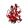 3roxC 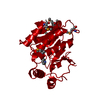 3rozC  3rphC  3rpzC  3rq5C  3rq6C 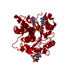 3rq8C 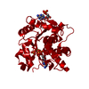 3rqhC  3rqqC  3rqxC  3rrbC  3rreC  3rrfC  3rrjC 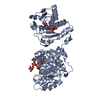 3rs8C 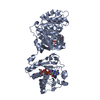 3rs9C  3rsfC 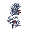 3rsgC  3rsqC  3rssC  3rt7C  3rt9C  3rtaC  3rtbC  3rtcC  3rtdC  3rteC  3rtgC  3ru2C  3ru3C  1kyhS S: Starting model for refinement C: citing same article ( |
|---|---|
| Similar structure data | |
| Other databases |


-Links
 PDBj
PDBj


- Assembly
| Deposited unit | 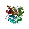
| ||||||||||||
|---|---|---|---|---|---|---|---|---|---|---|---|---|---|
| 1 | 
| ||||||||||||
| Unit cell |
| ||||||||||||
| Components on special symmetry positions |
|
-Components
| #1: Protein | Mass: 30176.439 Da / Num. of mol.: 1 Source method: isolated from a genetically manipulated source Source: (gene. exp.) Bacillus subtilis (bacteria) / Gene: BSU38720, yxkO / Plasmid: pMCSG19 / Production host: Escherichia coli (E. coli) / Strain (production host): BL21-CodonPlus (DE3)-RIPLReferences: UniProt: P94368, ATP-dependent NAD(P)H-hydrate dehydratase |
|---|---|
| #2: Chemical | ChemComp-MG /   Mass: 24.305 Da / Num. of mol.: 1 / Source method: obtained synthetically / Formula: Mg Mass: 24.305 Da / Num. of mol.: 1 / Source method: obtained synthetically / Formula: Mg |
| #3: Chemical | ChemComp-AMP / Adenosine monophosphate  Mass: 347.221 Da / Num. of mol.: 1 / Source method: obtained synthetically / Formula: C10H14N5O7P / Comment: AMP*YM Mass: 347.221 Da / Num. of mol.: 1 / Source method: obtained synthetically / Formula: C10H14N5O7P / Comment: AMP*YM |
| #4: Chemical | ChemComp-NAX /   Mass: 683.456 Da / Num. of mol.: 1 / Source method: obtained synthetically / Formula: C21H31N7O15P2 Mass: 683.456 Da / Num. of mol.: 1 / Source method: obtained synthetically / Formula: C21H31N7O15P2 |
| #5: Water | ChemComp-HOH / Water Mass: 18.015 Da / Num. of mol.: 184 / Source method: isolated from a natural source / Formula: H2O Mass: 18.015 Da / Num. of mol.: 184 / Source method: isolated from a natural source / Formula: H2O |
-Experimental details
-Experiment
| Experiment | Method: X-RAY DIFFRACTION / Number of used crystals: 1 |
|---|
- Sample preparation
| Crystal | Density Matthews: 2.97 Å3/Da / Density % sol: 58.57 % |
|---|---|
| Crystal grow | Temperature: 293 K / Method: vapor diffusion, hanging drop / pH: 7.5 Details: 0.005 M ATP, 0.18 M Magnesium Cloride, 13.5%(v/v) PEG 400, 10%(v/v) Glycerol, 0.09 M HEPES pH 7.5, VAPOR DIFFUSION, HANGING DROP, temperature 293K |
-Data collection
| Diffraction | Mean temperature: 100 K |
|---|---|
| Diffraction source | Source: SYNCHROTRON / Site: APS  / Beamline: 19-BM / Wavelength: 0.97918 Å / Beamline: 19-BM / Wavelength: 0.97918 Å |
| Detector | Type: ADSC QUANTUM 210r / Detector: CCD / Date: Oct 16, 2009 / Details: MIRRORS |
| Diffraction measurement | Details: 1.00 degrees, 1.00 sec, detector distance 160.00 mm |
| Radiation | Monochromator: SI(111) / Protocol: SINGLE WAVELENGTH / Monochromatic (M) / Laue (L): M / Scattering type: x-ray |
| Radiation wavelength | Wavelength: 0.97918 Å / Relative weight: 1 |
| Reflection | Av R equivalents: 0.083 / Number: 454188 |
| Reflection | Resolution: 1.8→50 Å / Num. obs: 33796 / % possible obs: 100 % / Observed criterion σ(F): 0 / Observed criterion σ(I): -3 / Redundancy: 13.4 % / Biso Wilson estimate: 31.5 Å2 / Rmerge(I) obs: 0.083 / Rsym value: 0.083 / Net I/σ(I): 45.786 |
| Reflection shell | Resolution: 1.8→1.83 Å / Redundancy: 11.8 % / Rmerge(I) obs: 0.896 / Mean I/σ(I) obs: 1.895 / Rsym value: 0.896 / % possible all: 99.9 |
| Cell measurement | Reflection used: 454188 |
- Processing
| Software |
| |||||||||||||||||||||||||||||||||||||||||||||||||||||||||||||||||||||||||||||||||||||
|---|---|---|---|---|---|---|---|---|---|---|---|---|---|---|---|---|---|---|---|---|---|---|---|---|---|---|---|---|---|---|---|---|---|---|---|---|---|---|---|---|---|---|---|---|---|---|---|---|---|---|---|---|---|---|---|---|---|---|---|---|---|---|---|---|---|---|---|---|---|---|---|---|---|---|---|---|---|---|---|---|---|---|---|---|---|---|
| Refinement | Method to determine structure: MOLECULAR REPLACEMENT Starting model: PDB ENTRY 1KYH Resolution: 1.8→50 Å / Cor.coef. Fo:Fc: 0.975 / Cor.coef. Fo:Fc free: 0.965 / Occupancy max: 1 / Occupancy min: 0.25 / SU B: 3.526 / SU ML: 0.059 / Cross valid method: THROUGHOUT / σ(F): 0 / ESU R Free: 0.089 / Stereochemistry target values: MAXIMUM LIKELIHOOD Details: HYDROGENS HAVE BEEN ADDED IN THE RIDING POSITIONS U VALUES : RESIDUAL ONLY
| |||||||||||||||||||||||||||||||||||||||||||||||||||||||||||||||||||||||||||||||||||||
| Solvent computation | Ion probe radii: 0.8 Å / Shrinkage radii: 0.8 Å / VDW probe radii: 1.2 Å / Solvent model: BABINET MODEL WITH MASK | |||||||||||||||||||||||||||||||||||||||||||||||||||||||||||||||||||||||||||||||||||||
| Displacement parameters | Biso max: 75.36 Å2 / Biso mean: 36.61 Å2 / Biso min: 25.48 Å2
| |||||||||||||||||||||||||||||||||||||||||||||||||||||||||||||||||||||||||||||||||||||
| Refinement step | Cycle: LAST / Resolution: 1.8→50 Å
| |||||||||||||||||||||||||||||||||||||||||||||||||||||||||||||||||||||||||||||||||||||
| Refine LS restraints |
| |||||||||||||||||||||||||||||||||||||||||||||||||||||||||||||||||||||||||||||||||||||
| LS refinement shell | Resolution: 1.804→1.851 Å / Total num. of bins used: 20
| |||||||||||||||||||||||||||||||||||||||||||||||||||||||||||||||||||||||||||||||||||||
| Refinement TLS params. | Method: refined / Origin x: -45.779 Å / Origin y: -21.241 Å / Origin z: -23.818 Å
|
