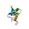
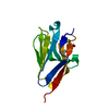
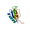
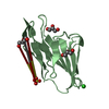
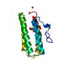
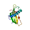
- PDB-6oqm: crystal structure of the MSH6 PWWP domain -
+
Open data
ID or keywords:
Loading...
-
Basic information
Entry
Database: PDB / ID: 6oqm
Title
crystal structure of the MSH6 PWWP domain
Components
DNA mismatch repair protein Msh6
Keywords
DNA BINDING PROTEIN / PWWP domain / Structural Genomics / Structural Genomics Consortium / SGC
Function / homology
Function and homology information
MutSalpha complex / Defective Mismatch Repair Associated With MSH2 / Defective Mismatch Repair Associated With MSH6 / guanine/thymine mispair binding / somatic recombination of immunoglobulin gene segments / positive regulation of helicase activity / meiotic mismatch repair / mismatched DNA binding / negative regulation of DNA recombination / Mismatch repair (MMR) directed by MSH2:MSH6 (MutSalpha) ...MutSalpha complex / Defective Mismatch Repair Associated With MSH2 / Defective Mismatch Repair Associated With MSH6 / guanine/thymine mispair binding / somatic recombination of immunoglobulin gene segments / positive regulation of helicase activity / meiotic mismatch repair / mismatched DNA binding / negative regulation of DNA recombination / Mismatch repair (MMR) directed by MSH2:MSH6 (MutSalpha) / isotype switching / ATP-dependent DNA damage sensor activity / mismatch repair / somatic hypermutation of immunoglobulin genes / ATP-dependent activity, acting on DNA / response to UV / methylated histone binding / intrinsic apoptotic signaling pathway / determination of adult lifespan / intrinsic apoptotic signaling pathway in response to DNA damage / spermatogenesis / damaged DNA binding / DNA repair / intracellular membrane-bounded organelle / chromatin binding / chromatin / Golgi apparatus / enzyme binding / nucleoplasm / ATP binding / nucleus / cytosol Similarity search - Function
DNA mismatch repair protein MutS/MSH / DNA mismatch repair protein MutS-like, N-terminal / DNA mismatch repair protein MutS, connector domain / DNA mismatch repair protein MutS, clamp / DNA mismatch repair protein MutS, N-terminal / MutS, connector domain superfamily / MutS domain I / MutS domain II / MutS family domain IV / MutS domain III ...DNA mismatch repair protein MutS/MSH / DNA mismatch repair protein MutS-like, N-terminal / DNA mismatch repair protein MutS, connector domain / DNA mismatch repair protein MutS, clamp / DNA mismatch repair protein MutS, N-terminal / MutS, connector domain superfamily / MutS domain I / MutS domain II / MutS family domain IV / MutS domain III / DNA mismatch repair MutS family / DNA mismatch repair protein MutS, C-terminal / DNA mismatch repair protein MutS, core / DNA mismatch repair protein MutS, core domain superfamily / MutS domain V / DNA mismatch repair proteins mutS family signature. / DNA-binding domain of DNA mismatch repair MUTS family / ATPase domain of DNA mismatch repair MUTS family / domain with conserved PWWP motif / PWWP domain / PWWP domain profile. / PWWP domain / P-loop containing nucleoside triphosphate hydrolase Similarity search - Domain/homology
In the structure databanks used in Yorodumi, some data are registered as the other names, "COVID-19 virus" and "2019-nCoV". Here are the details of the virus and the list of structure data.
Jan 31, 2019. EMDB accession codes are about to change! (news from PDBe EMDB page)
EMDB accession codes are about to change! (news from PDBe EMDB page)
The allocation of 4 digits for EMDB accession codes will soon come to an end. Whilst these codes will remain in use, new EMDB accession codes will include an additional digit and will expand incrementally as the available range of codes is exhausted. The current 4-digit format prefixed with “EMD-” (i.e. EMD-XXXX) will advance to a 5-digit format (i.e. EMD-XXXXX), and so on. It is currently estimated that the 4-digit codes will be depleted around Spring 2019, at which point the 5-digit format will come into force.
The EM Navigator/Yorodumi systems omit the EMD- prefix.
Related info.:Q: What is EMD? / ID/Accession-code notation in Yorodumi/EM Navigator
Yorodumi is a browser for structure data from EMDB, PDB, SASBDB, etc.
This page is also the successor to EM Navigator detail page, and also detail information page/front-end page for Omokage search.
The word "yorodu" (or yorozu) is an old Japanese word meaning "ten thousand". "mi" (miru) is to see.
Related info.:EMDB / PDB / SASBDB / Comparison of 3 databanks / Yorodumi Search / Aug 31, 2016. New EM Navigator & Yorodumi / Yorodumi Papers / Jmol/JSmol / Function and homology information / Changes in new EM Navigator and Yorodumi
 Movie
Movie Controller


 Open data
 Basic information
 Components
 Keywords
 Function and homology information
 Authors
 Citation
 Structure visualization
 Downloads & links
 Other downloads

 PDBj
PDBj

 Assembly
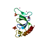


 Num. of mol.: 4 / Source method: obtained synthetically
 Mass: 24.305 Da / Num. of mol.: 1 / Source method: obtained synthetically / Formula: Mg
 Mass: 96.063 Da / Num. of mol.: 4 / Source method: obtained synthetically / Formula: SO4
 Mass: 18.015 Da / Num. of mol.: 2 / Source method: isolated from a natural source / Formula: H2O
 Sample preparation
 / Beamline: 19-ID / Wavelength: 0.9792 Å
 Processing