[English] 日本語
Yorodumi
- PDB-6ofs: The crystal structure of the periplasmic protease PqqL from Esche... -

+
Open data


ID or keywords:

Loading...

-
Basic information

Entry
Database: PDB / ID: 6ofs
TitleThe crystal structure of the periplasmic protease PqqL from Escherichia coli
ComponentsProbable zinc protease PqqL
KeywordsHYDROLASE / Protease / M16 Family / Processing Protease
Function / homology
Function and homology information


Hydrolases; Acting on peptide bonds (peptidases); Metalloendopeptidases / metalloendopeptidase activity / metallopeptidase activity / peptidase activity / outer membrane-bounded periplasmic space / proteolysis / zinc ion binding / cytosol
Similarity search - Function
Peptidase M16, zinc-binding site / Insulinase family, zinc-binding region signature. / Peptidase M16, C-terminal / Peptidase M16 inactive domain / Peptidase M16, N-terminal / Insulinase (Peptidase family M16) / Metalloenzyme, LuxS/M16 peptidase-like
Similarity search - Domain/homology
Probable zinc protease PqqL
Similarity search - Component
Biological speciesEscherichia coli (E. coli)
MethodX-RAY DIFFRACTION / SYNCHROTRON / MOLECULAR REPLACEMENT / Resolution: 2.6 Å
AuthorsGrinter, R.
Funding support United Kingdom, 1items
OrganizationGrant numberCountry
Wellcome Trust106077/Z/14/Z United Kingdom
Citation
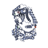
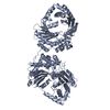
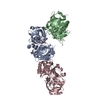
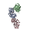
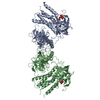
Journal: PLoS Genet / Year: 2019
Title: Protease-associated import systems are widespread in Gram-negative bacteria.
Authors: Rhys Grinter / Pok Man Leung / Lakshmi C Wijeyewickrema / Dene Littler / Simone Beckham / Robert N Pike / Daniel Walker / Chris Greening / Trevor Lithgow /
Abstract: Bacteria have evolved sophisticated uptake machineries in order to obtain the nutrients required for growth. Gram-negative plant pathogens of the genus Pectobacterium obtain iron from the protein ...Bacteria have evolved sophisticated uptake machineries in order to obtain the nutrients required for growth. Gram-negative plant pathogens of the genus Pectobacterium obtain iron from the protein ferredoxin, which is produced by their plant hosts. This iron-piracy is mediated by the ferredoxin uptake system (Fus), a gene cluster encoding proteins that transport ferredoxin into the bacterial cell and process it proteolytically. In this work we show that gene clusters related to the Fus are widespread in bacterial species. Through structural and biochemical characterisation of the distantly related Fus homologues YddB and PqqL from Escherichia coli, we show that these proteins are analogous to components of the Fus from Pectobacterium. The membrane protein YddB shares common structural features with the outer membrane ferredoxin transporter FusA, including a large extracellular substrate binding site. PqqL is an active protease with an analogous periplasmic localisation and iron-dependent expression to the ferredoxin processing protease FusC. Structural analysis demonstrates that PqqL and FusC share specific features that distinguish them from other members of the M16 protease family. Taken together, these data provide evidence that protease associated import systems analogous to the Fus are widespread in Gram-negative bacteria.
History
DepositionApr 1, 2019Deposition site: RCSB / Processing site: RCSB
Revision 1.0Oct 2, 2019Provider: repository / Type: Initial release
Revision 1.1Oct 30, 2019Group: Data collection / Database references / Category: citation / citation_author
Item: _citation.journal_volume / _citation.page_first ..._citation.journal_volume / _citation.page_first / _citation.page_last / _citation.pdbx_database_id_DOI / _citation.pdbx_database_id_PubMed / _citation.title
Revision 1.2Mar 13, 2024Group: Data collection / Database references / Category: chem_comp_atom / chem_comp_bond / database_2
Item: _database_2.pdbx_DOI / _database_2.pdbx_database_accession
Revision 1.3Apr 3, 2024Group: Refinement description / Category: pdbx_initial_refinement_model

-
Structure visualization

Structure viewerMolecule:
MolmilJmol/JSmol

Downloads & links

-
Assembly

Deposited unit
A: Probable zinc protease PqqL
hetero molecules


Theoretical massNumber of molelcules
Total (without water)103,0624
Polymers102,9261
Non-polymers1363
Water1,27971
1


  • Idetical with deposited unit
  • defined by author&software
  • Evidence: gel filtration
TypeNameSymmetry operationNumber
identity operation1_555x,y,z1
MethodPISA
Unit cell
Length a, b, c (Å)98.674, 98.674, 230.814
Angle α, β, γ (deg.)90.00, 90.00, 90.00
Int Tables number96
Space group name H-MP43212

-
Components

#1: Protein Probable zinc protease PqqL


Mass: 102925.570 Da / Num. of mol.: 1
Source method: isolated from a genetically manipulated source
Source: (gene. exp.) Escherichia coli (strain K12) (bacteria)
Strain: K12 / Gene: pqqL, yddC, b1494, JW1489 / Production host: Escherichia coli (E. coli)
References: UniProt: P31828, Hydrolases; Acting on peptide bonds (peptidases); Metalloendopeptidases
#2: Chemical ChemComp-ZN / ZINC ION


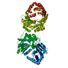
Mass: 65.409 Da / Num. of mol.: 1 / Source method: obtained synthetically / Formula: Zn
#3: Chemical ChemComp-CL / CHLORIDE ION / Chloride


Mass: 35.453 Da / Num. of mol.: 2 / Source method: obtained synthetically / Formula: Cl
#4: Water ChemComp-HOH / water / Water


Mass: 18.015 Da / Num. of mol.: 71 / Source method: isolated from a natural source / Formula: H2O

-
Experimental details

-
Experiment

ExperimentMethod: X-RAY DIFFRACTION / Number of used crystals: 1

-
Sample preparation

CrystalDensity Matthews: 2.73 Å3/Da / Density % sol: 54.93 %
Crystal growTemperature: 293 K / Method: evaporation / pH: 8.5
Details: 0.1 M Bis-tris propane, 0.2 M NaK tartrate, 20 % PEG 3350

-
Data collection

DiffractionMean temperature: 100 K / Serial crystal experiment: N
Diffraction sourceSource: SYNCHROTRON / Site: Australian Synchrotron / Beamline: MX1 / Wavelength: 0.987 Å
DetectorType: MAR CCD 130 mm / Detector: CCD / Date: Mar 8, 2016
RadiationMonochromator: Silicon Crystal / Protocol: SINGLE WAVELENGTH / Monochromatic (M) / Laue (L): M / Scattering type: x-ray
Radiation wavelengthWavelength: 0.987 Å / Relative weight: 1
ReflectionResolution: 2.6→48.25 Å / Num. obs: 36058 / % possible obs: 100 % / Observed criterion σ(I): 1.5 / Redundancy: 125.2 % / Biso Wilson estimate: 68.32 Å2 / CC1/2: 0.999 / Rmerge(I) obs: 0.354 / Rpim(I) all: 0.044 / Net I/σ(I): 25.1
Reflection shellResolution: 2.6→2.72 Å / Redundancy: 96.9 % / Rmerge(I) obs: 6.338 / Mean I/σ(I) obs: 1.6 / Num. unique obs: 4290 / CC1/2: 0.542 / Rpim(I) all: 0.645 / % possible all: 100

-
Processing

Software
NameVersionClassification
BUSTER2.10.3refinement
XDSdata reduction
Aimlessdata scaling
PHASERphasing
RefinementMethod to determine structure: MOLECULAR REPLACEMENT
Starting model: TBA

Resolution: 2.6→44.13 Å / Cor.coef. Fo:Fc: 0.945 / Cor.coef. Fo:Fc free: 0.931 / SU R Cruickshank DPI: 0.526 / Cross valid method: THROUGHOUT / σ(F): 0 / SU R Blow DPI: 0.533 / SU Rfree Blow DPI: 0.282 / SU Rfree Cruickshank DPI: 0.285
RfactorNum. reflection% reflectionSelection details
Rfree0.245 1737 4.83 %RANDOM
Rwork0.199 ---
obs0.201 35936 100 %-
Displacement parametersBiso mean: 64.04 Å2
Baniso -1Baniso -2Baniso -3
1-0.255 Å20 Å20 Å2
2--0.255 Å20 Å2
3----0.51 Å2
Refine analyzeLuzzati coordinate error obs: 0.35 Å
Refinement stepCycle: 1 / Resolution: 2.6→44.13 Å
ProteinNucleic acidLigandSolventTotal
Num. atoms7164 0 3 71 7238
Refine LS restraints
Refine-IDTypeDev idealNumberRestraint functionWeight
X-RAY DIFFRACTIONt_bond_d0.0097289HARMONIC2
X-RAY DIFFRACTIONt_angle_deg1.089891HARMONIC2
X-RAY DIFFRACTIONt_dihedral_angle_d2608SINUSOIDAL2
X-RAY DIFFRACTIONt_incorr_chiral_ct
X-RAY DIFFRACTIONt_pseud_angle
X-RAY DIFFRACTIONt_trig_c_planes
X-RAY DIFFRACTIONt_gen_planes1267HARMONIC5
X-RAY DIFFRACTIONt_it7289HARMONIC20
X-RAY DIFFRACTIONt_nbd0SEMIHARMONIC5
X-RAY DIFFRACTIONt_omega_torsion2.6
X-RAY DIFFRACTIONt_other_torsion19.77
X-RAY DIFFRACTIONt_improper_torsion
X-RAY DIFFRACTIONt_chiral_improper_torsion970SEMIHARMONIC5
X-RAY DIFFRACTIONt_sum_occupancies
X-RAY DIFFRACTIONt_utility_distance
X-RAY DIFFRACTIONt_utility_angle
X-RAY DIFFRACTIONt_utility_torsion
X-RAY DIFFRACTIONt_ideal_dist_contact8023SEMIHARMONIC4
LS refinement shellResolution: 2.6→2.62 Å / Total num. of bins used: 50
RfactorNum. reflection% reflection
Rfree0.32 -5.7 %
Rwork0.2297 678 -
all0.2346 719 -
obs--100 %
Refinement TLS params.Method: refined / Origin x: -41.2505 Å / Origin y: -10.9236 Å / Origin z: -13.0337 Å
111213212223313233
T0.0158 Å2-0.0938 Å20.0335 Å2--0.03 Å20.0699 Å2---0.0963 Å2
L0.1755 °2-0.0863 °20.0226 °2-0.5353 °20.3645 °2--0.1622 °2
S0.0577 Å °-0.0406 Å °-0.0535 Å °0.058 Å °-0.0605 Å °-0.0453 Å °-0.0096 Å °0.0139 Å °0.0028 Å °
Refinement TLS groupSelection details: { A|* }

+
About Yorodumi

-
News

-
Feb 9, 2022. New format data for meta-information of EMDB entries

New format data for meta-information of EMDB entries

  • Version 3 of the EMDB header file is now the official format.
  • The previous official version 1.9 will be removed from the archive.

Related info.:EMDB header

External links:wwPDB to switch to version 3 of the EMDB data model

-
Aug 12, 2020. Covid-19 info

Covid-19 info

URL: https://pdbj.org/emnavi/covid19.php

New page: Covid-19 featured information page in EM Navigator.

Related info.:Covid-19 info / Mar 5, 2020. Novel coronavirus structure data

+
Mar 5, 2020. Novel coronavirus structure data

Novel coronavirus structure data

Related info.:Yorodumi Speices / Aug 12, 2020. Covid-19 info

External links:COVID-19 featured content - PDBj / Molecule of the Month (242):Coronavirus Proteases

+
Jan 31, 2019. EMDB accession codes are about to change! (news from PDBe EMDB page)

EMDB accession codes are about to change! (news from PDBe EMDB page)

  • The allocation of 4 digits for EMDB accession codes will soon come to an end. Whilst these codes will remain in use, new EMDB accession codes will include an additional digit and will expand incrementally as the available range of codes is exhausted. The current 4-digit format prefixed with “EMD-” (i.e. EMD-XXXX) will advance to a 5-digit format (i.e. EMD-XXXXX), and so on. It is currently estimated that the 4-digit codes will be depleted around Spring 2019, at which point the 5-digit format will come into force.
  • The EM Navigator/Yorodumi systems omit the EMD- prefix.

Related info.:Q: What is EMD? / ID/Accession-code notation in Yorodumi/EM Navigator

External links:EMDB Accession Codes are Changing Soon! / Contact to PDBj

+
Jul 12, 2017. Major update of PDB

Major update of PDB

  • wwPDB released updated PDB data conforming to the new PDBx/mmCIF dictionary.
  • This is a major update changing the version number from 4 to 5, and with Remediation, in which all the entries are updated.
  • In this update, many items about electron microscopy experimental information are reorganized (e.g. em_software).
  • Now, EM Navigator and Yorodumi are based on the updated data.

External links:wwPDB Remediation / Enriched Model Files Conforming to OneDep Data Standards Now Available in the PDB FTP Archive

-
Yorodumi

Thousand views of thousand structures

  • Yorodumi is a browser for structure data from EMDB, PDB, SASBDB, etc.
  • This page is also the successor to EM Navigator detail page, and also detail information page/front-end page for Omokage search.
  • The word "yorodu" (or yorozu) is an old Japanese word meaning "ten thousand". "mi" (miru) is to see.

Related info.:EMDB / PDB / SASBDB / Comparison of 3 databanks / Yorodumi Search / Aug 31, 2016. New EM Navigator & Yorodumi / Yorodumi Papers / Jmol/JSmol / Function and homology information / Changes in new EM Navigator and Yorodumi

Read more