 Movie
Movie Controller

[English] 日本語
 Yorodumi
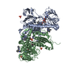
- PDB-6oex: Crystal structure of Trypanothione Reductase from Trypanosoma bru... -
+ Open data

- Basic information
| Entry | Database: PDB / ID: 6oex | ||||||
|---|---|---|---|---|---|---|---|
| Title | Crystal structure of Trypanothione Reductase from Trypanosoma brucei in complex with inhibitor 3-(2-{1-[2-(Piperidin-4-yl)ethyl]-1H-indol-5-yl}-5-[1-(pyrrolidin-1-yl)cyclohexyl]-1,3- thiazol-4-yl)-N-(2,2,2-trifluoroethyl)prop-2-yn-1-amine | ||||||
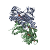
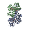
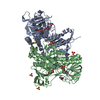
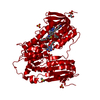
 Components Components | Trypanothione reductase | ||||||
 Keywords Keywords | oxidoreductase/oxidoreductase inhibitor /  Trypanosoma / trypanothione / inhibitor / sleeping sickness / OXIDOREDUCTASE / oxidoreductase-oxidoreductase inhibitor complex Trypanosoma / trypanothione / inhibitor / sleeping sickness / OXIDOREDUCTASE / oxidoreductase-oxidoreductase inhibitor complex | ||||||
| Function / homology |  Function and homology informationtrypanothione-disulfide reductase / trypanothione-disulfide reductase (NADPH) activity / glutathione-disulfide reductase (NADPH) activity / glycosome / thioredoxin-disulfide reductase (NADPH) activity / ciliary plasm / glutathione metabolic process / cell redox homeostasis / cellular response to oxidative stress / flavin adenine dinucleotide binding ...trypanothione-disulfide reductase / trypanothione-disulfide reductase (NADPH) activity / glutathione-disulfide reductase (NADPH) activity / glycosome / thioredoxin-disulfide reductase (NADPH) activity / ciliary plasm / glutathione metabolic process / cell redox homeostasis / cellular response to oxidative stress / flavin adenine dinucleotide binding / mitochondrion / nucleoplasm / metal ion binding / cytosol / cytoplasm Function and homology informationtrypanothione-disulfide reductase / trypanothione-disulfide reductase (NADPH) activity / glutathione-disulfide reductase (NADPH) activity / glycosome / thioredoxin-disulfide reductase (NADPH) activity / ciliary plasm / glutathione metabolic process / cell redox homeostasis / cellular response to oxidative stress / flavin adenine dinucleotide binding ...trypanothione-disulfide reductase / trypanothione-disulfide reductase (NADPH) activity / glutathione-disulfide reductase (NADPH) activity / glycosome / thioredoxin-disulfide reductase (NADPH) activity / ciliary plasm / glutathione metabolic process / cell redox homeostasis / cellular response to oxidative stress / flavin adenine dinucleotide binding / mitochondrion / nucleoplasm / metal ion binding / cytosol / cytoplasmSimilarity search - Function | ||||||
| Biological species |  Trypanosoma brucei brucei (eukaryote) Trypanosoma brucei brucei (eukaryote) | ||||||
| Method | X-RAY DIFFRACTION / SYNCHROTRON / MOLECULAR REPLACEMENT / Resolution: 2.1 Å | ||||||
 Authors Authors | Halgas, O. / De Gasparo, R. / Harangozo, D. / Krauth-Siegel, R.L. / Diederich, F. / Pai, E.F. | ||||||
| Funding support |  Canada, 1items Canada, 1items
| ||||||
 Citation Citation | Journal: Chemistry / Year: 2019 Title: Targeting a Large Active Site: Structure-Based Design of Nanomolar Inhibitors of Trypanosoma brucei Trypanothione Reductase. Authors: De Gasparo, R. / Halgas, O. / Harangozo, D. / Kaiser, M. / Pai, E.F. / Krauth-Siegel, R.L. / Diederich, F. | ||||||
| History |
|
- Structure visualization
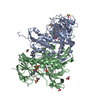
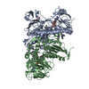
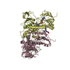
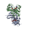
| Structure viewer | Molecule: MolmilJmol/JSmol |
|---|
- Downloads & links
-Download
| PDBx/mmCIF format | 6oex.cif.gz | 417.5 KB | Display | PDBx/mmCIF format |
|---|---|---|---|---|
| PDB format | pdb6oex.ent.gz | 339.5 KB | Display | PDB format |
| PDBx/mmJSON format | 6oex.json.gz | Tree view | PDBx/mmJSON format | |
| Others |  Other downloads Other downloads |
-Validation report
| Arichive directory | https://data.pdbj.org/pub/pdb/validation_reports/oe/6oexftp://data.pdbj.org/pub/pdb/validation_reports/oe/6oex | HTTPS FTP |
|---|
-Related structure data
| Related structure data |  6oeyC  6oezC  4nevS S: Starting model for refinement C: citing same article ( |
|---|---|
| Similar structure data |


-Links
 PDBj
PDBj


- Assembly
| Deposited unit | 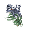
| ||||||||
|---|---|---|---|---|---|---|---|---|---|
| 1 |
| ||||||||
| Unit cell |
|
-Components
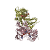
-Protein , 1 types, 2 molecules BA
| #1: Protein | Mass: 53497.969 Da / Num. of mol.: 2 Source method: isolated from a genetically manipulated source Source: (gene. exp.) Trypanosoma brucei brucei (strain 927/4 GUTat10.1) (eukaryote)Strain: 927/4 GUTat10.1 / Gene: Tb10.406.0520 / Plasmid: PET3ATBTRYR Production host:  Escherichia coli 'BL21-Gold(DE3)pLysS AG' (bacteria) Escherichia coli 'BL21-Gold(DE3)pLysS AG' (bacteria)References: UniProt: Q389T8, trypanothione-disulfide reductase |
|---|
-Non-polymers , 5 types, 571 molecules 


| #2: Chemical | Flavin adenine dinucleotide Mass: 785.550 Da / Num. of mol.: 2 Mass: 785.550 Da / Num. of mol.: 2Source method: isolated from a genetically manipulated source Formula: C27H33N9O15P2 / Comment: FAD*YM #3: Chemical | HEPES Mass: 238.305 Da / Num. of mol.: 2 / Source method: obtained synthetically / Formula: C8H18N2O4S / Comment: pH buffer*YM Mass: 238.305 Da / Num. of mol.: 2 / Source method: obtained synthetically / Formula: C8H18N2O4S / Comment: pH buffer*YM#4: Chemical | Sulfate Mass: 96.063 Da / Num. of mol.: 2 / Source method: obtained synthetically / Formula: SO4 Mass: 96.063 Da / Num. of mol.: 2 / Source method: obtained synthetically / Formula: SO4#5: Chemical |  Mass: 597.780 Da / Num. of mol.: 2 / Source method: obtained synthetically / Formula: C33H42F3N5S Mass: 597.780 Da / Num. of mol.: 2 / Source method: obtained synthetically / Formula: C33H42F3N5S#6: Water | ChemComp-HOH / | WaterMass: 18.015 Da / Num. of mol.: 563 / Source method: isolated from a natural source / Formula: H2O |
|---|
-Experimental details
-Experiment
| Experiment | Method: X-RAY DIFFRACTION / Number of used crystals: 1 |
|---|
- Sample preparation
| Crystal | Density Matthews: 3.6 Å3/Da / Density % sol: 65.8 % / Description: yellowish tetragonal bipyramid |
|---|---|
| Crystal grow | Temperature: 298 K / Method: vapor diffusion, hanging drop / pH: 7.5 Details: 2 microL of protein were mixed with 2 microL of well solution (500 microL; 0.1 M HEPES, pH 7.5, 2.2 M (NH4)2SO4). Crystals grew within 2 weeks. |
-Data collection
| Diffraction | Mean temperature: 100 K / Serial crystal experiment: N |
|---|---|
| Diffraction source | Source: SYNCHROTRON / Site: APS  / Beamline: 17-ID / Wavelength: 1 Å / Beamline: 17-ID / Wavelength: 1 Å |
| Detector | Type: DECTRIS PILATUS 6M / Detector: PIXEL / Date: Aug 12, 2018 |
| Radiation | Protocol: SINGLE WAVELENGTH / Monochromatic (M) / Laue (L): M / Scattering type: x-ray |
| Radiation wavelength | Wavelength: 1 Å / Relative weight: 1 |
| Reflection | Resolution: 2.1→46.4 Å / Num. obs: 90546 / % possible obs: 96 % / Redundancy: 13.2 % / CC1/2: 0.999 / Net I/σ(I): 16.9 |
| Reflection shell | Resolution: 2.1→2.18 Å / Num. unique obs: 7051 / CC1/2: 0.637 |
- Processing
| Software |
| |||||||||||||||||||||||||||||||||||||||||||||||||||||||||||||||||||||||||||||||||||||||||||||||||||||||||||||||||||||||||||||||||||||||||||||||||||||||||||||||||||||||||||||||||||||||||||||||||||||||||||||||||||||||||
|---|---|---|---|---|---|---|---|---|---|---|---|---|---|---|---|---|---|---|---|---|---|---|---|---|---|---|---|---|---|---|---|---|---|---|---|---|---|---|---|---|---|---|---|---|---|---|---|---|---|---|---|---|---|---|---|---|---|---|---|---|---|---|---|---|---|---|---|---|---|---|---|---|---|---|---|---|---|---|---|---|---|---|---|---|---|---|---|---|---|---|---|---|---|---|---|---|---|---|---|---|---|---|---|---|---|---|---|---|---|---|---|---|---|---|---|---|---|---|---|---|---|---|---|---|---|---|---|---|---|---|---|---|---|---|---|---|---|---|---|---|---|---|---|---|---|---|---|---|---|---|---|---|---|---|---|---|---|---|---|---|---|---|---|---|---|---|---|---|---|---|---|---|---|---|---|---|---|---|---|---|---|---|---|---|---|---|---|---|---|---|---|---|---|---|---|---|---|---|---|---|---|---|---|---|---|---|---|---|---|---|---|---|---|---|---|---|---|---|
| Refinement | Method to determine structure: MOLECULAR REPLACEMENT Starting model: 4NEV Resolution: 2.1→46.4 Å / SU ML: 0.18 / Cross valid method: FREE R-VALUE / σ(F): 1.34 / Phase error: 19.19
| |||||||||||||||||||||||||||||||||||||||||||||||||||||||||||||||||||||||||||||||||||||||||||||||||||||||||||||||||||||||||||||||||||||||||||||||||||||||||||||||||||||||||||||||||||||||||||||||||||||||||||||||||||||||||
| Solvent computation | Shrinkage radii: 0.9 Å / VDW probe radii: 1.11 Å | |||||||||||||||||||||||||||||||||||||||||||||||||||||||||||||||||||||||||||||||||||||||||||||||||||||||||||||||||||||||||||||||||||||||||||||||||||||||||||||||||||||||||||||||||||||||||||||||||||||||||||||||||||||||||
| Refinement step | Cycle: LAST / Resolution: 2.1→46.4 Å
| |||||||||||||||||||||||||||||||||||||||||||||||||||||||||||||||||||||||||||||||||||||||||||||||||||||||||||||||||||||||||||||||||||||||||||||||||||||||||||||||||||||||||||||||||||||||||||||||||||||||||||||||||||||||||
| Refine LS restraints |
| |||||||||||||||||||||||||||||||||||||||||||||||||||||||||||||||||||||||||||||||||||||||||||||||||||||||||||||||||||||||||||||||||||||||||||||||||||||||||||||||||||||||||||||||||||||||||||||||||||||||||||||||||||||||||
| LS refinement shell |
| |||||||||||||||||||||||||||||||||||||||||||||||||||||||||||||||||||||||||||||||||||||||||||||||||||||||||||||||||||||||||||||||||||||||||||||||||||||||||||||||||||||||||||||||||||||||||||||||||||||||||||||||||||||||||
| Refinement TLS params. | Method: refined / Origin x: -12.4489 Å / Origin y: -31.9354 Å / Origin z: -49.5888 Å
| |||||||||||||||||||||||||||||||||||||||||||||||||||||||||||||||||||||||||||||||||||||||||||||||||||||||||||||||||||||||||||||||||||||||||||||||||||||||||||||||||||||||||||||||||||||||||||||||||||||||||||||||||||||||||
| Refinement TLS group | Selection details: all |
