 Movie
Movie Controller


+ Open data

- Basic information
| Entry | Database: PDB / ID: 2wov | ||||||
|---|---|---|---|---|---|---|---|
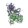
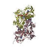
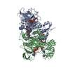
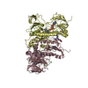
| Title | Trypanosoma brucei trypanothione reductase with bound NADP. | ||||||
 Components Components | TRYPANOTHIONE REDUCTASE | ||||||
 Keywords Keywords |  OXIDOREDUCTASE / TRYPANOSOMIASIS / SLEEPING SICKNESS / FLAVOPROTEIN / TRYPANOTHIONE / REDUCTASE / REDOX-ACTIVE CENTER OXIDOREDUCTASE / TRYPANOSOMIASIS / SLEEPING SICKNESS / FLAVOPROTEIN / TRYPANOTHIONE / REDUCTASE / REDOX-ACTIVE CENTER | ||||||
| Function / homology |  Function and homology informationtrypanothione-disulfide reductase / trypanothione-disulfide reductase (NADPH) activity / glutathione-disulfide reductase (NADPH) activity / glycosome / thioredoxin-disulfide reductase (NADPH) activity / ciliary plasm / glutathione metabolic process / cell redox homeostasis / cellular response to oxidative stress / flavin adenine dinucleotide binding ...trypanothione-disulfide reductase / trypanothione-disulfide reductase (NADPH) activity / glutathione-disulfide reductase (NADPH) activity / glycosome / thioredoxin-disulfide reductase (NADPH) activity / ciliary plasm / glutathione metabolic process / cell redox homeostasis / cellular response to oxidative stress / flavin adenine dinucleotide binding / mitochondrion / nucleoplasm / metal ion binding / cytosol / cytoplasm Function and homology informationtrypanothione-disulfide reductase / trypanothione-disulfide reductase (NADPH) activity / glutathione-disulfide reductase (NADPH) activity / glycosome / thioredoxin-disulfide reductase (NADPH) activity / ciliary plasm / glutathione metabolic process / cell redox homeostasis / cellular response to oxidative stress / flavin adenine dinucleotide binding ...trypanothione-disulfide reductase / trypanothione-disulfide reductase (NADPH) activity / glutathione-disulfide reductase (NADPH) activity / glycosome / thioredoxin-disulfide reductase (NADPH) activity / ciliary plasm / glutathione metabolic process / cell redox homeostasis / cellular response to oxidative stress / flavin adenine dinucleotide binding / mitochondrion / nucleoplasm / metal ion binding / cytosol / cytoplasmSimilarity search - Function | ||||||
| Biological species |  TRYPANOSOMA BRUCEI (eukaryote) TRYPANOSOMA BRUCEI (eukaryote) | ||||||
| Method | X-RAY DIFFRACTION / MOLECULAR REPLACEMENT / Resolution: 2.5 Å | ||||||
 Authors Authors | Alphey, M.S. / Fairlamb, A.H. | ||||||
 Citation Citation | Journal: J.Med.Chem. / Year: 2011 Title: Dihydroquinazolines as a Novel Class of Trypanosoma Brucei Trypanothione Reductase Inhibitors: Discovery, Synthesis, and Characterization of Their Binding Mode by Protein Crystallography. Authors: Patterson, S. / Alphey, M.S. / Jones, D.C. / Shanks, E.J. / Street, I.P. / Frearson, J.A. / Wyatt, P.G. / Gilbert, I.H. / Fairlamb, A.H. | ||||||
| History |
| ||||||
| Remark 700 | SHEET THE SHEET STRUCTURE OF THIS MOLECULE IS BIFURCATED. IN ORDER TO REPRESENT THIS FEATURE IN ... SHEET THE SHEET STRUCTURE OF THIS MOLECULE IS BIFURCATED. IN ORDER TO REPRESENT THIS FEATURE IN THE SHEET RECORDS BELOW, TWO SHEETS ARE DEFINED. |
- Structure visualization
| Structure viewer | Molecule: MolmilJmol/JSmol |
|---|
- Downloads & links
-Download
| PDBx/mmCIF format | 2wov.cif.gz | 393.2 KB | Display | PDBx/mmCIF format |
|---|---|---|---|---|
| PDB format | pdb2wov.ent.gz | 320.1 KB | Display | PDB format |
| PDBx/mmJSON format | 2wov.json.gz | Tree view | PDBx/mmJSON format | |
| Others |  Other downloads Other downloads |
-Validation report
| Arichive directory | https://data.pdbj.org/pub/pdb/validation_reports/wo/2wovftp://data.pdbj.org/pub/pdb/validation_reports/wo/2wov | HTTPS FTP |
|---|
-Related structure data
| Related structure data |  2woiSC  2wowC  2wp5C  2wp6C  2wpcC  2wpeC  2wpfC S: Starting model for refinement C: citing same article ( |
|---|---|
| Similar structure data |


-Links
 PDBj
PDBj


- Assembly
| Deposited unit | 
| ||||||||||||||||
|---|---|---|---|---|---|---|---|---|---|---|---|---|---|---|---|---|---|
| 1 | 
| ||||||||||||||||
| 2 | 
| ||||||||||||||||
| Unit cell |
| ||||||||||||||||
| Noncrystallographic symmetry (NCS) | NCS oper:
|
-Components
| #1: Protein | Mass: 53497.969 Da / Num. of mol.: 4 Source method: isolated from a genetically manipulated source Source: (gene. exp.) TRYPANOSOMA BRUCEI (eukaryote) / Strain: TREU927 / Plasmid: PET15B / Production host:  ESCHERICHIA COLI (E. coli) / Strain (production host): BL21(DE3) / Variant (production host): CODONPLUS RIL ESCHERICHIA COLI (E. coli) / Strain (production host): BL21(DE3) / Variant (production host): CODONPLUS RILReferences: UniProt: Q389T8, trypanothione-disulfide reductase#2: Chemical | ChemComp-NDP / Nicotinamide adenine dinucleotide phosphate  Mass: 745.421 Da / Num. of mol.: 4 / Source method: obtained synthetically / Formula: C21H30N7O17P3 Mass: 745.421 Da / Num. of mol.: 4 / Source method: obtained synthetically / Formula: C21H30N7O17P3#3: Chemical | ChemComp-FAD / Flavin adenine dinucleotide  Mass: 785.550 Da / Num. of mol.: 4 / Source method: obtained synthetically / Formula: C27H33N9O15P2 / Comment: FAD*YM Mass: 785.550 Da / Num. of mol.: 4 / Source method: obtained synthetically / Formula: C27H33N9O15P2 / Comment: FAD*YM#4: Chemical | ChemComp-NA /   Mass: 22.990 Da / Num. of mol.: 7 / Source method: obtained synthetically / Formula: Na Mass: 22.990 Da / Num. of mol.: 7 / Source method: obtained synthetically / Formula: Na#5: Water | ChemComp-HOH / | Water Mass: 18.015 Da / Num. of mol.: 440 / Source method: isolated from a natural source / Formula: H2O Mass: 18.015 Da / Num. of mol.: 440 / Source method: isolated from a natural source / Formula: H2O |
|---|
-Experimental details
-Experiment
| Experiment | Method: X-RAY DIFFRACTION / Number of used crystals: 1 |
|---|
- Sample preparation
| Crystal | Density Matthews: 2.52 Å3/Da / Density % sol: 51.2 % / Description: NONE |
|---|---|
| Crystal grow | Details: 15MG/ML PROTEIN IN 25MM HEPES PH 7.5 AND 50MM NABR EQUILIBRATED AGAINST 24% MPD, 10% PEG3350 AND 40MM IMIDAZOLE PH 8.0. |
-Data collection
| Diffraction | Mean temperature: 100 K |
|---|---|
| Diffraction source | Source: ROTATING ANODE / Type: RIGAKU MICROMAX-007 / Wavelength: 1.5418 |
| Detector | Type: RIGAKU RAXIS IV / Detector: IMAGE PLATE / Date: Jan 13, 2009 / Details: MIRRORS |
| Radiation | Protocol: SINGLE WAVELENGTH / Monochromatic (M) / Laue (L): M / Scattering type: x-ray |
| Radiation wavelength | Wavelength: 1.5418 Å / Relative weight: 1 |
| Reflection | Resolution: 2.5→19.9 Å / Num. obs: 72546 / % possible obs: 93.3 % / Observed criterion σ(I): 2 / Redundancy: 3.6 % / Biso Wilson estimate: 37 Å2 / Rmerge(I) obs: 0.11 / Net I/σ(I): 14.8 |
| Reflection shell | Resolution: 2.5→2.56 Å / Redundancy: 3.5 % / Rmerge(I) obs: 0.4 / Mean I/σ(I) obs: 3.6 / % possible all: 85.6 |
- Processing
| Software |
| ||||||||||||||||||||||||||||||||||||||||||||||||||||||||||||||||||||||||||||||||||||||||||||||||||||||||||||||||||||||||||||||||||||||||||||||||||||||||||||||||||||||||||||||||||||||
|---|---|---|---|---|---|---|---|---|---|---|---|---|---|---|---|---|---|---|---|---|---|---|---|---|---|---|---|---|---|---|---|---|---|---|---|---|---|---|---|---|---|---|---|---|---|---|---|---|---|---|---|---|---|---|---|---|---|---|---|---|---|---|---|---|---|---|---|---|---|---|---|---|---|---|---|---|---|---|---|---|---|---|---|---|---|---|---|---|---|---|---|---|---|---|---|---|---|---|---|---|---|---|---|---|---|---|---|---|---|---|---|---|---|---|---|---|---|---|---|---|---|---|---|---|---|---|---|---|---|---|---|---|---|---|---|---|---|---|---|---|---|---|---|---|---|---|---|---|---|---|---|---|---|---|---|---|---|---|---|---|---|---|---|---|---|---|---|---|---|---|---|---|---|---|---|---|---|---|---|---|---|---|---|
| Refinement | Method to determine structure: MOLECULAR REPLACEMENT Starting model: PDB ENTRY 2WOI Resolution: 2.5→19.847 Å / Cor.coef. Fo:Fc: 0.946 / Cor.coef. Fo:Fc free: 0.891 / SU B: 9.955 / SU ML: 0.223 / Cross valid method: THROUGHOUT / ESU R: 0.73 / ESU R Free: 0.304 / Stereochemistry target values: MAXIMUM LIKELIHOOD Details: HYDROGENS HAVE BEEN ADDED IN THE RIDING POSITIONS. SOME RESIDUES AT N- AND C-TERMINI WERE MODELLED AS ALANINES OR OMITTED FROM THE MODEL DUE TO THEIR FLEXIBILITY AND POOR ELECTRON DENSITY
| ||||||||||||||||||||||||||||||||||||||||||||||||||||||||||||||||||||||||||||||||||||||||||||||||||||||||||||||||||||||||||||||||||||||||||||||||||||||||||||||||||||||||||||||||||||||
| Solvent computation | Ion probe radii: 0.8 Å / VDW probe radii: 1.4 Å / Solvent model: MASK BULK SOLVENT | ||||||||||||||||||||||||||||||||||||||||||||||||||||||||||||||||||||||||||||||||||||||||||||||||||||||||||||||||||||||||||||||||||||||||||||||||||||||||||||||||||||||||||||||||||||||
| Displacement parameters | Biso mean: 27.915 Å2
| ||||||||||||||||||||||||||||||||||||||||||||||||||||||||||||||||||||||||||||||||||||||||||||||||||||||||||||||||||||||||||||||||||||||||||||||||||||||||||||||||||||||||||||||||||||||
| Refinement step | Cycle: LAST / Resolution: 2.5→19.847 Å
| ||||||||||||||||||||||||||||||||||||||||||||||||||||||||||||||||||||||||||||||||||||||||||||||||||||||||||||||||||||||||||||||||||||||||||||||||||||||||||||||||||||||||||||||||||||||
| Refine LS restraints |
|